

The PQ-loop protein Any1 segregates Drs2 and Neo1 functions required for viability and plasma membrane phospholipid asymmetry
- PMID: 30824614
- PMCID: PMC6495175
- DOI: 10.1194/jlr.M093526
The PQ-loop protein Any1 segregates Drs2 and Neo1 functions required for viability and plasma membrane phospholipid asymmetry
Abstract
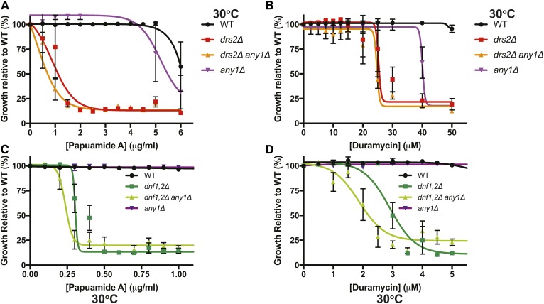
Membrane asymmetry is a key organizational feature of the plasma membrane. Type IV P-type ATPases (P4-ATPases) are phospholipid flippases that establish membrane asymmetry by translocating phospholipids, such as phosphatidylserine (PS) and phospatidylethanolamine, from the exofacial leaflet to the cytosolic leaflet. Saccharomyces cerevisiae expresses five P4-ATPases: Drs2, Neo1, Dnf1, Dnf2, and Dnf3. The inactivation of Neo1 is lethal, suggesting Neo1 mediates an essential function not exerted by the other P4-ATPases. However, the disruption of ANY1, which encodes a PQ-loop membrane protein, allows the growth of neo1Δ and reveals functional redundancy between Golgi-localized Neo1 and Drs2. Here we show Drs2 PS flippase activity is required to support neo1Δ any1Δ viability. Additionally, a Dnf1 variant with enhanced PS flipping ability can replace Drs2 and Neo1 function in any1Δ cells. any1Δ also suppresses drs2Δ growth defects but not the loss of membrane asymmetry. Any1 overexpression perturbs the growth of cells but does not disrupt membrane asymmetry. Any1 coimmunoprecipitates with Neo1, an association prevented by the Any1-inactivating mutation D84G. These results indicate a critical role for PS flippase activity in Golgi membranes to sustain viability and suggests Any1 regulates Golgi membrane remodeling through protein-protein interactions rather than a previously proposed scramblase activity.
Keywords: Golgi apparatus; phosphatidylcholine; phosphatidylethanolamine; phosphatidylserine; trafficking; transport.
Copyright © 2019 Takar et al.
Figures


Similar articles
-
Conserved mechanism of phospholipid substrate recognition by the P4-ATPase Neo1 from Saccharomyces cerevisiae.Biochim Biophys Acta Mol Cell Biol Lipids. 2020 Feb;1865(2):158581. doi: 10.1016/j.bbalip.2019.158581. Epub 2019 Nov 28. Biochim Biophys Acta Mol Cell Biol Lipids. 2020. PMID: 31786280 Free PMC article.
-
The Essential Neo1 Protein from Budding Yeast Plays a Role in Establishing Aminophospholipid Asymmetry of the Plasma Membrane.J Biol Chem. 2016 Jul 22;291(30):15727-39. doi: 10.1074/jbc.M115.686253. Epub 2016 May 26. J Biol Chem. 2016. PMID: 27235400 Free PMC article.
-
Identification of residues defining phospholipid flippase substrate specificity of type IV P-type ATPases.Proc Natl Acad Sci U S A. 2012 Feb 7;109(6):E290-8. doi: 10.1073/pnas.1115725109. Epub 2012 Jan 20. Proc Natl Acad Sci U S A. 2012. PMID: 22308393 Free PMC article.
-
Substrates of P4-ATPases: beyond aminophospholipids (phosphatidylserine and phosphatidylethanolamine).FASEB J. 2019 Mar;33(3):3087-3096. doi: 10.1096/fj.201801873R. Epub 2018 Dec 3. FASEB J. 2019. PMID: 30509129 Review.
-
Functions of phospholipid flippases.J Biochem. 2011 Feb;149(2):131-43. doi: 10.1093/jb/mvq140. Epub 2010 Dec 5. J Biochem. 2011. PMID: 21134888 Review.
Cited by
-
A complex genetic interaction implicates that phospholipid asymmetry and phosphate homeostasis regulate Golgi functions.PLoS One. 2020 Jul 30;15(7):e0236520. doi: 10.1371/journal.pone.0236520. eCollection 2020. PLoS One. 2020. PMID: 32730286 Free PMC article.
-
Genome-wide CRISPR screen reveals CLPTM1L as a lipid scramblase required for efficient glycosylphosphatidylinositol biosynthesis.Proc Natl Acad Sci U S A. 2022 Apr 5;119(14):e2115083119. doi: 10.1073/pnas.2115083119. Epub 2022 Mar 28. Proc Natl Acad Sci U S A. 2022. PMID: 35344438 Free PMC article.
-
Conserved mechanism of phospholipid substrate recognition by the P4-ATPase Neo1 from Saccharomyces cerevisiae.Biochim Biophys Acta Mol Cell Biol Lipids. 2020 Feb;1865(2):158581. doi: 10.1016/j.bbalip.2019.158581. Epub 2019 Nov 28. Biochim Biophys Acta Mol Cell Biol Lipids. 2020. PMID: 31786280 Free PMC article.
-
Structural basis of the P4B ATPase lipid flippase activity.Nat Commun. 2021 Oct 13;12(1):5963. doi: 10.1038/s41467-021-26273-0. Nat Commun. 2021. PMID: 34645814 Free PMC article.
-
Functional unknomics: Systematic screening of conserved genes of unknown function.PLoS Biol. 2023 Aug 8;21(8):e3002222. doi: 10.1371/journal.pbio.3002222. eCollection 2023 Aug. PLoS Biol. 2023. PMID: 37552676 Free PMC article.
References
-
- Gordesky S. E., Marinetti G. V., and Love R.. 1975. The reaction of chemical probes with the erythrocyte membrane. J. Membr. Biol. 20: 111–132. - PubMed
-
- Verkleij A. J., Zwaal R. F., Roelofsen B., Comfurius P., Kastelijn D., and van Deenen L. L.. 1973. The asymmetric distribution of phospholipids in the human red cell membrane. A combined study using phospholipases and freeze-etch electron microscopy. Biochim. Biophys. Acta. 323: 178–193. - PubMed
-
- Bretscher M. S. 1972. Asymmetrical lipid bilayer structure for biological membranes. Nat. New Biol. 236: 11–12. - PubMed
-
- Daleke D. L. 2003. Regulation of transbilayer plasma membrane phospholipid asymmetry. J. Lipid Res. 44: 233–242. - PubMed
-
- López-Marques R. L., Poulsen L. R., Bailly A., Geisler M., Pomorski T. G., and Palmgren M. G.. 2015. Structure and mechanism of ATP-dependent phospholipid transporters. Biochim. Biophys. Acta. 1850: 461–475. - PubMed
Publication types
MeSH terms
Substances
Grants and funding
LinkOut - more resources
Full Text Sources
Molecular Biology Databases
