Observational Study
. 2019 Mar 1;9(1):3289.
doi: 10.1038/s41598-019-39772-4.

Mobilization of Stem and Progenitor Cells in Septic Shock Patients

Affiliations
Observational Study

Mobilization of Stem and Progenitor Cells in Septic Shock Patients

Tomasz Skirecki et al. Sci Rep. .

Abstract

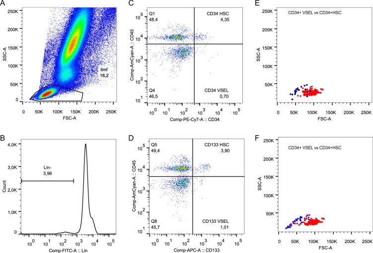
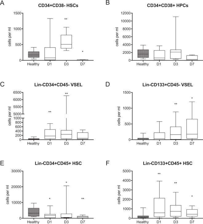
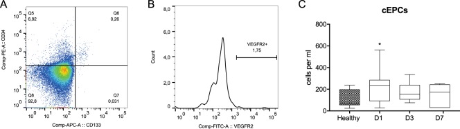
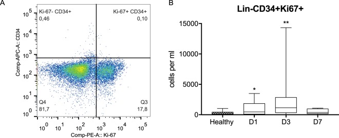
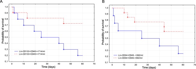
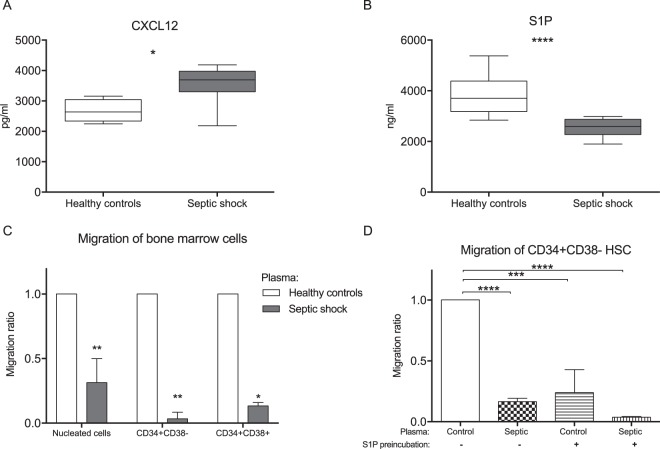
Septic shock is associated with multiple injuries to organs and tissues. These events may induce the regenerative response of adult stem cells. However, little is known about how endogenous stem cells are modulated by sepsis. This study analyzed the circulation of hematopoietic stem cells (HSCs), endothelial progenitor cells (EPCs) and very small embryonic-like stem cells (VSELs) in the peripheral blood of patients with septic shock. Thirty-three patients with septic shock and twenty-two healthy control subjects were enrolled in this prospective observational study. Blood samples were collected on the first, third and seventh days of septic shock. Populations of stem cells were analyzed by flow cytometry. Chemotactic mediators were analyzed by HPLC and ELISA. Populations of early HSCs (Lin-CD133+CD45+ and CD34+CD38-) were mobilized to the peripheral blood after an initial decrease. Mobilized HSCs showed significantly increased expression of Ki-67, a marker of cell proliferation. Circulating EPCs and VSELs were mobilized to the blood circulation upon the first day of sepsis. Patients with a greater number of Lin-CD133+CD45+ HSCs and Lin-CD34+CD45- VSELs had a significantly lower probability of 60-day survival. The concentration of CXCL12 was elevated in the blood of septic patients, while the concentration of sphingosine-1-phosphate was significantly decreased. As an emergency early response to sepsis, VSELs and EPCs were mobilized to the peripheral blood, while the HSCs showed delayed mobilization. Differential mobilization of stem cell subsets reflected changes in the concentration of chemoattractants in the blood. The relationship between the probability of death and a large number of HSCs and VSELs in septic shock patients can be used as a novel prognostic marker and may provide new therapeutic approaches.


Conflict of interest statement

The authors declare no competing interests.

Figures

Figure 1
Figure 1
Representative gating strategy for the analysis of VSELs and HSCs. (A) First, an extended lymphocyte gate was created, (B) then cells negative for the Lineage cocktail antigens are gated. (C) Lin- cells were then analyzed for the expression of CD45 and CD34 and (D) CD45 and CD133. (E) Lin-CD34+CD45+ HSCs and Lin-CD34+CD45− VSELs were then plotted on the SSC/FSC dot plot to compare their granularity and size. (F) SSC/FSC dot plot graph showing Lin-CD133+CD45− VSELs and Lin-CD133+CD45+ HSCs.
Figure 2
Figure 2
Total numbers of circulating stem cell subpopulations per mL of peripheral blood. (A) CD34+CD38– HSCs. (B) CD34+CD38+ HPCs. (C) Lin-CD34+CD45− VSELs. (D) Lin-CD133+CD45− VSELs. (E) Lin-CD34+CD45+ HSCs. (F) Lin-CD133+CD45+ HSCs. Comparisons between the control and septic groups were performed by Mann-Whitney U test. Statistical significance between values for healthy controls and septic shock patients are marked as follows: *p < 0.05 and **p < 0.01. The comparison of the cell number between days 1 and 3 by the Wilcoxon signed-rank test did not reveal significant differences.
Figure 3
Figure 3
Analysis of the circulating endothelial cells. (A) A cytogram of CD34 versus CD133 expression gated from the extended lymphocyte gate (Fig. 1A). (B) VEGFR2 receptor expression by double-positive progenitor cells. (C) Dynamics of the changes in the total number of cEPCs in septic shock patients. Cell counts between healthy controls and septic shock patients were compared using Mann-Whitney U test and the statistical significance was marked as follows: *p < 0.05. Cell counts between days 1 and 3 was compared by the Wilcoxon signed-rank test which did not reveal significant differences.
Figure 4
Figure 4
Analysis of the proliferation of circulating hematopoietic stem and progenitor cells by the expression of Ki-67. (A) Lineage-negative cells (Fig. 1A,B) were analyzed for the expression of the Ki-67 antigen in the CD34+ cells. (B) The dynamics of the circulation of Lin-CD34+Ki-67+ cells in septic shock patients. Cell counts between healthy controls and septic shock patients were compared using Mann-Whitney U test and the statistical significance was marked as follows: *p < 0.05. Cell counts between days 1 and 3 was compared by the Wilcoxon signed-rank test which did not reveal significant differences.
Figure 5
Figure 5
Probability of 60-day survival of septic shock patients in relation to the number of circulating stem cells. (A) Survival curves of patients with varied number of Lin-CD133+CD45+ HSCs. (B) Survival curves of patients with varied numbers of Lin-CD34+CD45− VSELs.
Figure 6
Figure 6
Mechanisms of stem cell mobilization. (A) Serum levels of CXCL12 in septic shock patients and healthy volunteers. (B) Plasma concentrations of S1P in septic shock and in controls. (C) The chemotactic activity of septic plasma was evaluated using a Transwell migration assay on human bone marrow cells. The migration of all nucleated cells, CD34+CD38− HSCs and CD34+CD38+ HPCs was analyzed. The results were normalized to the number of cells that migrated to the normal human plasma (n = 5). (D) Migration of bone marrow cells that were preincubated in 100 nM S1P or medium only for 60 minutes before the chemotaxis assay. The concentration of mediators was compared using Mann-Whitney U test (A,B) and comparisons between multiple groups were performed using Anova with Dunnet’s multiple comparison test (C,D) *p < 0.05, **p < 0.01, ***p < 0.001 and ****p < 0.0001.

References

    1. Levy MM, et al. Outcomes of the Surviving Sepsis Campaign in intensive care units in the USA and Europe: a prospective cohort study. Lancet Infect Dis. 2012;12:919–924. doi: 10.1016/S1473-3099(12)70239-6. - DOI - PubMed
    1. Abraham E, Singer M. Mechanisms of sepsis-induced organ dysfunction. Crit Care Med. 2007;35:2408–2416. doi: 10.1097/01.CCM.0000282072.56245.91. - DOI - PubMed
    1. Manz MG, Boettcher S. Emergency granulopoiesis. Nat Rev Immunol. 2014;14:302–314. doi: 10.1038/nri3660. - DOI - PubMed
    1. Skirecki T, et al. Early severe impairment of hematopoietic stem and progenitor cells from the bone marrow caused by CLP sepsis and endotoxemia in a humanized mice model. Stem Cell Res Ther. 2015;6:142. doi: 10.1186/s13287-015-0135-9. - DOI - PMC - PubMed
    1. Rodriguez S, et al. Dysfunctional expansion of hematopoietic stem cells and block of myeloid differentiation in lethal sepsis. Blood. 2009;114:4064–4076. doi: 10.1182/blood-2009-04-214916. - DOI - PMC - PubMed

Publication types