. 2019 Mar 1;9(1):3248.
doi: 10.1038/s41598-019-39011-w.

Designing a whole cell bioreporter to show antioxidant activities of agents that work by promotion of the KEAP1-NRF2 signaling pathway

Affiliations

Designing a whole cell bioreporter to show antioxidant activities of agents that work by promotion of the KEAP1-NRF2 signaling pathway

Negar Mozaheb et al. Sci Rep. .

Erratum in

Abstract

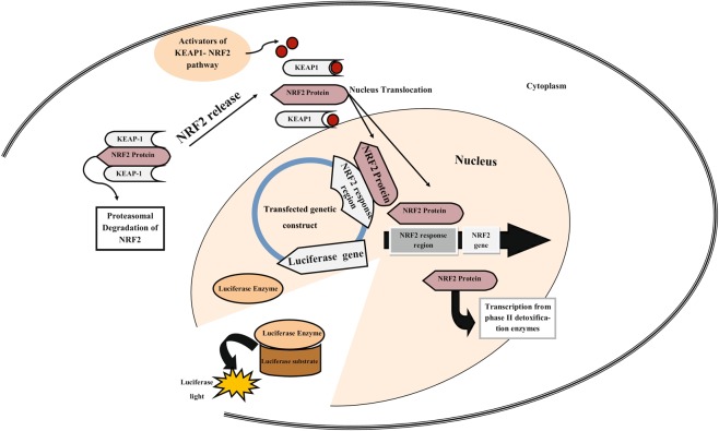
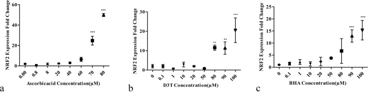
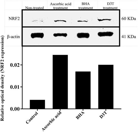
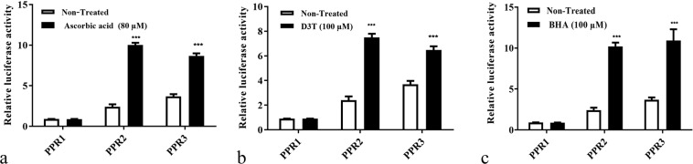
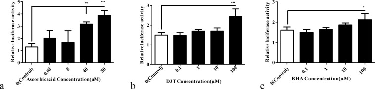
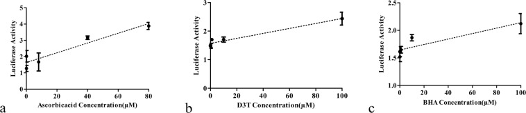
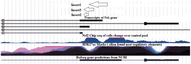
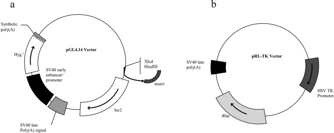
The major signaling pathway in human cells is related to the antioxidant defense system. The main component of this system is a transcription factor, Nuclear Factor Erythroid 2-Related Factor 2 (NRF2). It regulates this system in different cellular situations under stimulation by oxidative stress or antioxidants. Thus, detecting the stimulation of NRF2 via a screening strategy may enable us to discover stimulating agents of NRF2-related signaling pathway. With this in mind, we designed a whole cell bioreporter containing the NRF2 response elements that are inserted in a luciferase vector, immediately upstream of a luciferase gene whose promoter has been removed. This bioreporter is activated by stimulators such as 3H-1,2-dithiole-3-thione (D3T), butyl hydroxyanisole (BHA) and ascorbic acid reacting as antioxidant agents. It was observed that the regulatory region of the NRF2 gene, which is identified by NRF2 protein, is located inside its coding region. This designed bioreporter can detect the presence of antioxidant agents. It also exhibits a significant linear correlation over different doses of these agents ranging from 0.8 to 80 μM for ascorbic acid, 0.1 to 100 μM for D3T, and 0.1 to 100 μM for BHA. This detection system is proven to be more sensitive than Real-time PCR, suggesting it to be a highly sensitive system among the available methods.


Conflict of interest statement

The authors declare no competing interests.

Figures

Figure 1
Figure 1
Schematic of our bioreporter. During NRF2-KEAP1 pathway activation, NRF2 is disassociated from its repressor protein in the cytoplasm, KEAP1, followed by its translocation to the nucleus, binding to its ARE at the upstream of the NRF2 gene and also that of transfected vector’s luciferase.
Figure 2
Figure 2
Effects of different concentrations of antioxidants on NRF2 expression. The meaningful overexpression of the NRF2 at concentrations of about 80, 90 and 100 µM of ascorbic acid (a), 80, 90 and 100 µM of D3T (b), and 90 and 100 µM of BHA (c). Values reported are the average ± SD of three independent experiments. ***P-value < 0.001, **P-value < 0.01 using One-way ANOVA data analyzing.
Figure 3
Figure 3
Western blot analysis shows an increase in NRF2 protein level in cells. The cells were treated with ascorbic acid (80 µM), BHA (100 µM), and D3T (100 µM) and after 12 hours the NRF2 protein level was compared to non-treated control. The graphs are related to optical density of NRF2 bands that are normalized by the internal control (β-actin). Full-length blots with molecular marker are presented in Supplementary Fig. S1.
Figure 4
Figure 4
Comparative graph showing effects of maximum concentrations of antioxidants, (a) ascorbic acid (80 µM), (b) D3T (100 µM), and (c) BHA (100 µM) on three transfected HEK 293 cells with different genetic constructs, using Eq. 1. Reported values are the average ± SD of three independent experiments and they indicate better induction of PPR2 when affected by ascorbic acid. ***P-value < 0.001 by Two- way ANOVA data analyzing.
Figure 5
Figure 5
Effects of different concentrations of antioxidants on the activity of NRF2 response region, using Eq. 1. HEK 293 cells were transfected with PPR2 and then treated with (a) 0.8, 8, 40 and 80 μM of ascorbic acid, (b) 0.1, 1, 10 and 100 μM of D3T, and (c); 0.1, 1, 10 and 100 μM of BHA. Reported values are the average ± SD of the three independent experiments. ***P-value < 0.001, **P-value < 0.01, and *P-value < 0.5 by One-way ANOVA data analyzing.
Figure 6
Figure 6
The luciferase activity of bioreporter against different concentrations of antioxidants (a) ascorbic acid (0.8, 8, 40 and 80 μM), (b) D3T (0.1, 1, 10 and 100 μM), and (c) BHA (0.1, 1, 10 and 100 μM). The results show a linear relationship between luciferase activity and doses of the antioxidants.
Figure 7
Figure 7
Image of UCSC output. Insert 1, Insert 2 and Insert 3 are probable promoter regions including those that were cloned.
Figure 8
Figure 8
Vector maps. (a) pGL4.14 vector, (b) pRL-TK vector.

References

    1. Sies H. Oxidative stress: a concept in redox biology and medicine. Redox Biol. 2015;4:180–183. doi: 10.1016/j.redox.2015.01.002. - DOI - PMC - PubMed
    1. Obulesu M, Dowlathabad MR, Bramhachari P. Carotenoids and Alzheimer’s disease: an insight into therapeutic role of retinoids in animal models. Neurochem. Int. 2011;59:535–541. doi: 10.1016/j.neuint.2011.04.004. - DOI - PubMed
    1. Kubo E, Chhunchha B, Singh P, Sasaki H, Singh DP. Sulforaphane reactivates cellular antioxidant defense by inducing Nrf2/ARE/Prdx6 activity during aging and oxidative stress. Sci. Rep. 2017;7:14130. doi: 10.1038/s41598-017-14520-8. - DOI - PMC - PubMed
    1. Reddy SP. The antioxidant response element and oxidative stress modifiers in airway diseases. Curr. Mol. Med. 2008;8:376–383. doi: 10.2174/156652408785160925. - DOI - PMC - PubMed
    1. Akcakaya H, Tok S, Dal F, Cinar SA, Nurten R. beta-carotene treatment alters the cellular death process in oxidative stress-induced K562 cells. Cell Biol Int. 2017;41:309–319. doi: 10.1002/cbin.10727. - DOI - PubMed