. 2019 Jun:32:28-37.
doi: 10.1016/j.gep.2019.02.001. Epub 2019 Feb 27.

Expression of the heparin-binding growth factors Midkine and pleiotrophin during ocular development

Affiliations

Expression of the heparin-binding growth factors Midkine and pleiotrophin during ocular development

Ruda Cui et al. Gene Expr Patterns. 2019 Jun.

Abstract

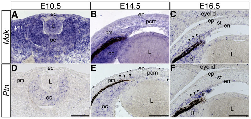
Midkine (MDK) and Pleiotrophin (PTN) belong to a group of heparin-binding growth factors that has been shown to have pleiotropic functions in various biological processes during development and disease. Development of the vertebrate eye is a multistep process that involves coordinated interactions between neuronal and non-neuronal cells, but very little is known about the potential function of MDK and PTN in these processes. In this study, we demonstrate by section in situ hybridization, the spatiotemporal expression of MDK and PTN during ocular development in chick and mouse. We show that MDK and PTN are expressed in dynamic patterns that overlap in a few non-neuronal tissues in the anterior eye and in neuronal cell layers of the posterior eye. We show that the expression patterns of MDK and PTN are only conserved in a few tissues in chick and mouse but they overlap with the expression of some of their receptors LRP1, RPTPZ, ALK, NOTCH2, ITGβ1, SDC1, and SDC3. The dynamic expression patterns of MDK, PTN and their receptors suggest that they function together during the multistep process of ocular development and they may play important roles in cell proliferation, adhesion, and migration of neuronal and non-neuronal cells.

Keywords: Cornea; Lens; Midkine; Ocular development; Pleiotrophin; Retina.


Figures

Figure 1.
Figure 1.. Expression pattern of MDK and PTN during development of the chick anterior eye.
Transverse sections through E3, E5, and E7 eyes showing the spatiotemporal expression of MDK (A-C) and PTN (D-F). Asterisks in B, C, E, and F indicate ocular blood vessels. Arrowheads in F indicate condensed mesenchyme at the boundary between the presumptive drainage angle and trabecular meshwork. Abbreviations: ec, ectoderm; pm, periocular mesenchyme; oc, optic cup; L, lens; ep, corneal epithelium; en, corneal endothelium; st, corneal stroma; ir, iris; cm, ciliary margin. Scale bars represent 100 μm in A, D; B, E; and C, F.
Figure 2.
Figure 2.. Expression pattern of Mdk and Ptn during development of the mouse anterior eye.
Transverse sections through E10.5, E14.5, and E16.5 eyes showing the spatiotemporal expression of Mdk (A-C) and Ptn (D-F). Arrowheads in E and F indicate expression of Ptn in mesenchyme of the presumptive iris. Abbreviations: ec, ectoderm; pm, periocular mesenchyme; oc, optic cup; L, lens; ep, corneal epithelium; pcm, presumptive corneal mesenchyme; en, corneal endothelium; st, corneal stroma; ir, iris; pm, periocular mesenchyme; R, neural retina. Scale bars represent 100 μm in A, D; B, E; and C, F.
Figure 3.
Figure 3.. Expression pattern of MDK and PTN during retinal development in chick.
Transverse sections through E5, E7, E10, E15 and E18 eyes showing the spatiotemporal expression of MDK (A-E) and PTN (F-J). All images oriented with the retinal pigmented epithelium (rpe) at the top. Images for A and F represent posterior retina. Rest of the images were acquired from the dorsal retina in the region between the optic nerve and ciliary body. Asterisks in B and G indicate detachment of the rpe (not shown) from the neural retina during tissue processing. Abbreviations: nr, neural retina; mes, mesenchyme; on, optic nerve; gcl, ganglion cell layer; inl, inner nuclear layer; onl, outer nuclear layer. Scale bars represent 100 μm in A,F and B,C,D,E,G,H,I,J.
Figure 4.
Figure 4.. Expression pattern of Mdk and Ptn during retinal development in mouse.
Transverse sections through E13.5, E14.5, E16.5, P0 and P12 eyes showing the spatiotemporal expression of Mdk (A-E) and Ptn (F-J). All images oriented with the retinal pigmented epithelium (rpe) at the top. Images for A and F represent the posterior retina. Rest of the images were acquired from the dorsal retina in the region between the optic nerve and ciliary body. Abbreviations: mes, mesenchyme; on, optic nerve; onbl, outer neuroblastic layer; inbl, inner neuroblastic layer; inl, inner nuclear layer; gcl, ganglion cell layer. Scale bar represents 100 μm.
Figure 5.
Figure 5.. Expression of MDK and PTN receptors during chick ocular development.
(A-D) Transverse sections through E7 anterior eye showing the expression of NOTCH2, ITGβ1, SDC1, and SDC3. (E-K) Transverse sections through E7 retina showing the expression of NOTCH2, ITGβ1, SDC1, SDC3, LRP1, RPTPZ, and ALK. (L) RTPCR results showing amplification of NOTCH2, ITGβ1, LRP1, RPTPZ, ALK, SDC1, SDC3, and GAPDH (control) using cDNA obtained from E7 chick corneas and retinas. All retina images were acquired from the dorsal retina in the region between the optic nerve and ciliary body. Abbreviations: ep, corneal epithelium; st, corneal stroma; en, corneal endothelium; ir, iris; L, lens; rpe, retinal pigmented epithelium; gcl, ganglion cell layer; inl, inner nuclear layer; C, cornea; R, retina. Arrowheads indicate condensed mesenchyme that marks the boundary between the presumptive drainage angle and trabecular meshwork. Asterisks in A and C indicate ocular blood vessels. Scale bar represents 100 μm in A,B,C,D; and 100μm in E,F,G,H,I,J,K.
Figure 6.
Figure 6.. Expression pattern of MDK and PTN in the anterior eye of E12 chick and receptor expression in the trigeminal ganglion.
Transverse sections through E12 anterior eye showing the expression of (A) MDK in mostly epithelial tissues (arrowheads) and (B) PTN in mesenchymal tissues. (C) RTPCR results showing amplification of NOTCH2, ITGβ1, LRP1, RPTPZ, ALK, SDC1, SDC3, and GAPDH (control) using cDNA obtained from E12 chick trigeminal ganglia. Cross-sections through E12 trigeminal ganglion showing the expression of ITGβ1 (D, D’) and RPTPZ (E, E’), and MDK (F, F’) and PTN (G, G’). Asterisk in A indicates ocular blood vessel..Abbreviations: nm, nictitating membrane; ir, iris; cb, ciliary body; ica, iridocorneal angle; L, lens; m, ciliary muscle; opV, ophthalmic branch of the trigeminal ganglion; mmV, maxillomandibular branch of the trigeminal ganglion. Scale bars represent 250μm in A,B; 250μm in D,E,F,G; and 100μm in D’,E’,F’,G’.

Similar articles

Cited by

References

    1. Agathocleous M, Harris WA, 2009. From Progenitors to Differentiated Cells in the Vertebrate Retina. Annu Rev Cell Dev Biol 25, 45–69. 10.1146/annurev.cellbio.042308.113259 - DOI - PubMed
    1. Asahina K, Sato H, Yamasaki C, Kataoka M, Shiokawa M, Katayama S, Tateno C, Yoshizato K, 2002. Pleiotrophin/heparin-binding growth-associated molecule as a mitogen of rat hepatocytes and its role in regeneration and development of liver. Am. J. Pathol. 160, 2191–2205. 10.1016/S0002-9440(10)61167-4 - DOI - PMC - PubMed
    1. Beebe DC, Coats JM, 2000. The Lens Organizes the Anterior Segment: Specification of Neural Crest Cell Differentiation in the Avian Eye. Dev. Biol. 220, 424–431. 10.1006/dbio.2000.9638 - DOI - PubMed
    1. Calinescu AA, Vihtelic TS, Hyde DR, Hitchcock PF, 2009. Cellular expression of midkine-a and midkine-b during retinal development and photoreceptor regeneration in zebrafish. J. Comp. Neurol. 514, 1–10. 10.1002/cne.21999 - DOI - PMC - PubMed
    1. Canoll PD, Barnea G, Levy JB, Sap J, Ehrlich M, Silvennoinen O, Schlessinger J, Musacchio J, 1993. The expression of a novel receptor-type tyrosine phosphatase suggests a role in morphogenesis and plasticity of the nervous system. Dev. Brain Res. 75, 293–298. 10.1016/0165-3806(93)90035-9 - DOI - PubMed

Publication types

MeSH terms