. 2019 May 16;26(5):662-673.e7.
doi: 10.1016/j.chembiol.2019.01.012. Epub 2019 Feb 28.

Characterization, Dynamics, and Mechanism of CXCR4 Antagonists on a Constitutively Active Mutant

Affiliations

Characterization, Dynamics, and Mechanism of CXCR4 Antagonists on a Constitutively Active Mutant

Eric M Rosenberg Jr et al. Cell Chem Biol. .

Abstract

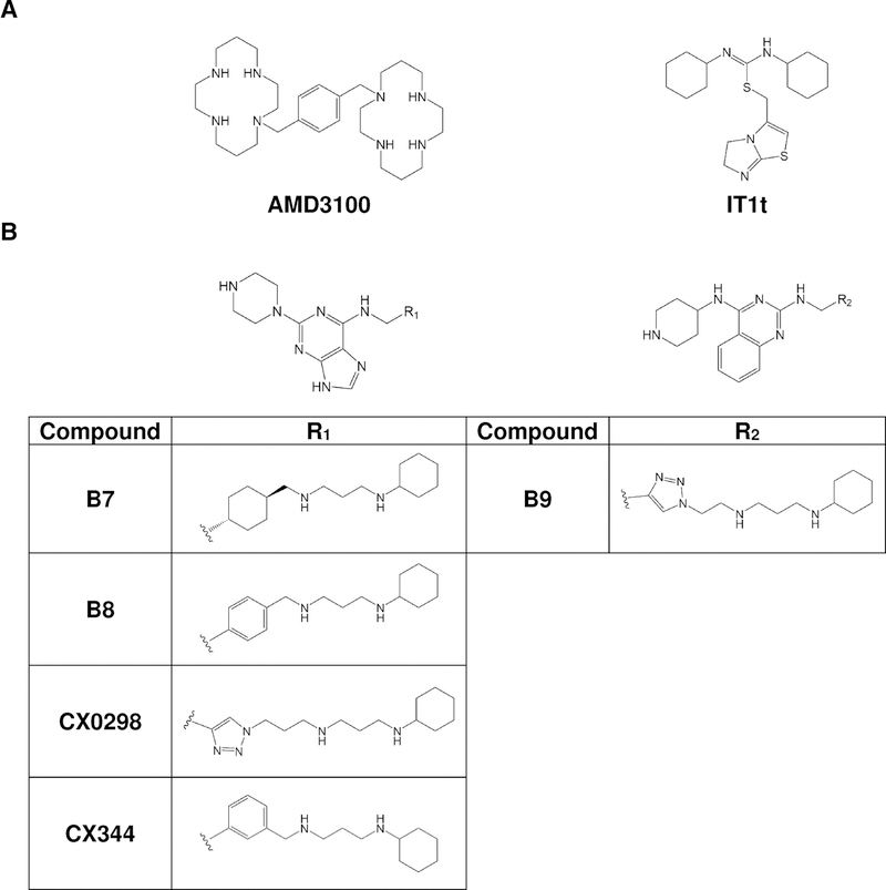
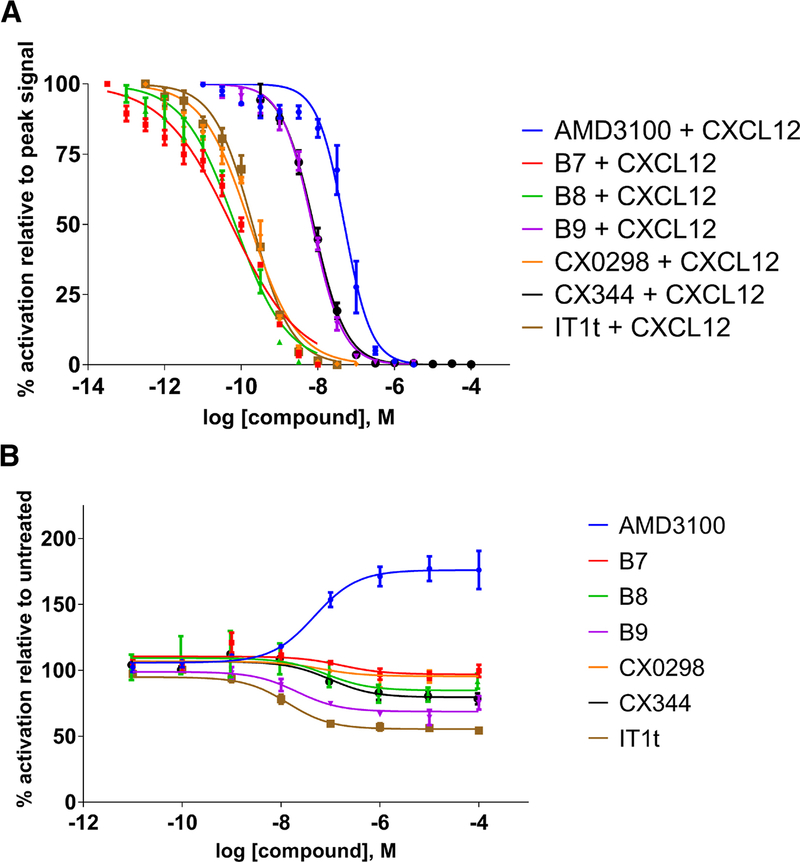
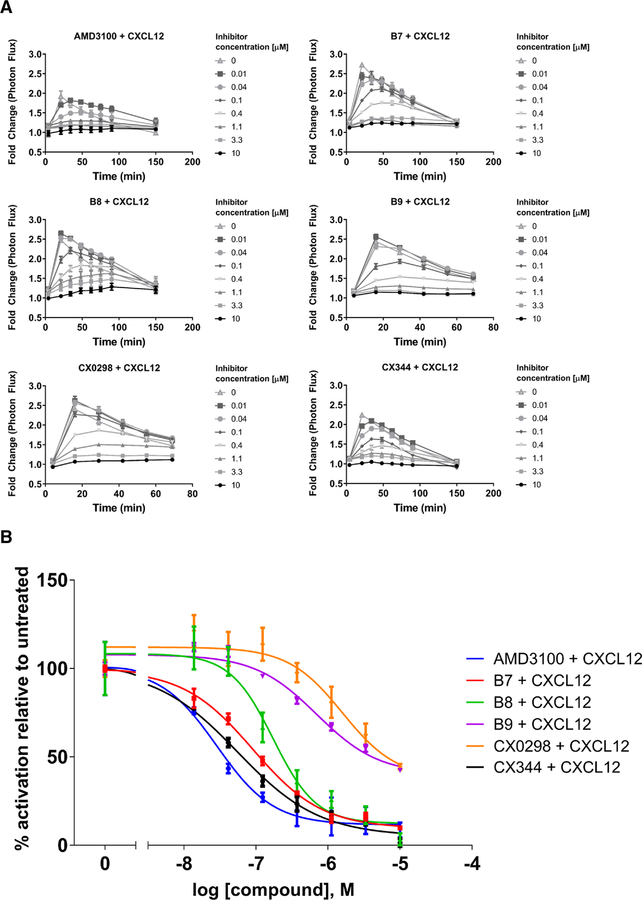
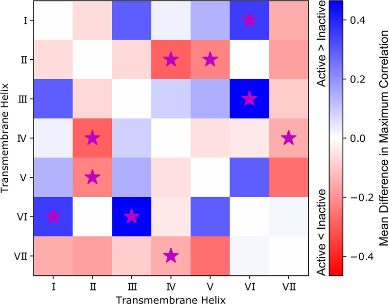
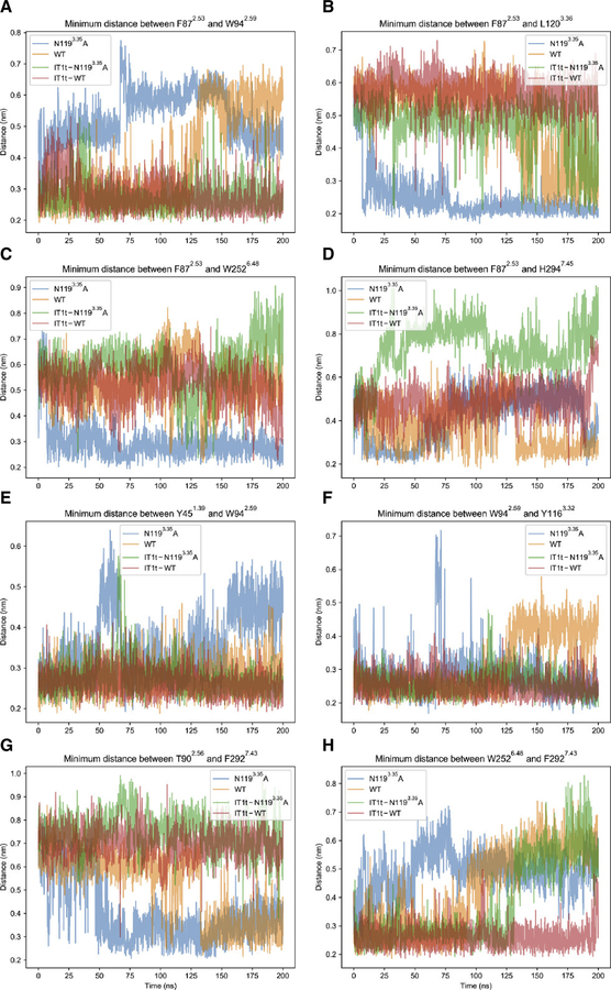
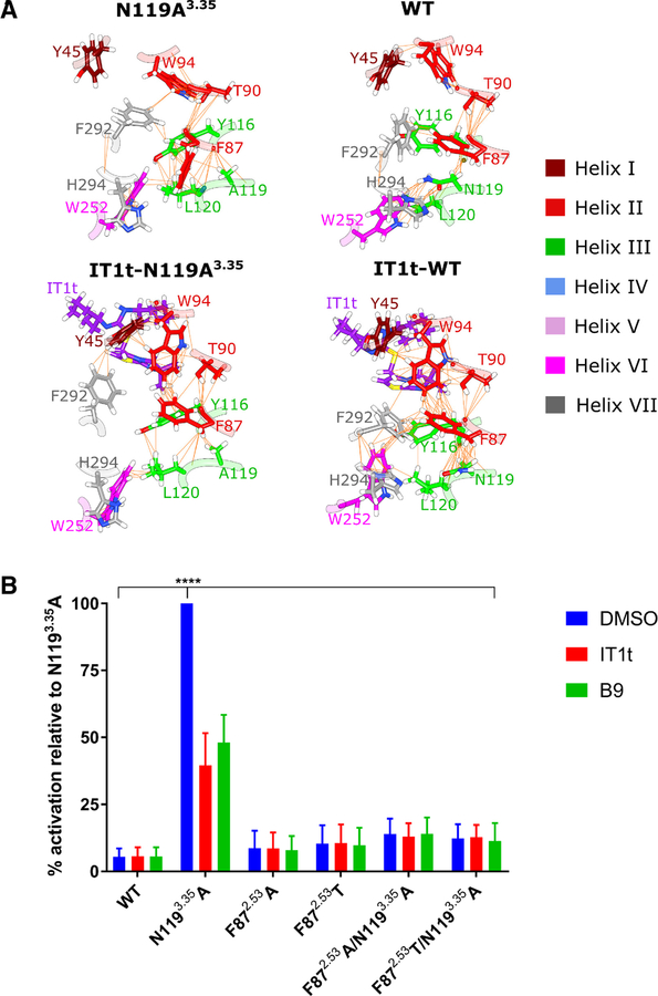
The G protein-coupled receptor (GPCR) CXCR4 is a co-receptor for HIV and is involved in cancers and autoimmune diseases. We characterized five purine or quinazoline core polyamine pharmacophores used for targeting CXCR4 dysregulation in diseases. All were neutral antagonists for wild-type CXCR4 and two were biased antagonists with effects on β-arrestin-2 only at high concentrations. These compounds displayed various activities for a constitutively active mutant (CAM). We use the IT1t-CXCR4 crystal structure and molecular dynamics (MD) simulations to develop two hypotheses for the activation of the N1193.35A CAM. The N1193.35A mutation facilitates increased coupling of TM helices III and VI. IT1t deactivates the CAM by disrupting the coupling between TM helices III and VI, mediated primarily by residue F872.53. Mutants of F872.53 in N1193.35A CXCR4 precluded constitutive signaling and prevented inverse agonism. This work characterizes CXCR4 ligands and provides a mechanism for N1193.35A constitutive activation.

Keywords: CXCL12; CXCR4; G protein-coupled receptor (GPCR); constitutively active mutant (CAM); molecular dynamics (MD); small molecule ligands.


Conflict of interest statement

DECLARATION OF INTERESTS

KEL and GDL receive research funding from Polyphor.

Figures

FIGURE 1.
FIGURE 1.
A. Structures of the FDA-approved drug AMD3100 and the small molecule antagonist IT1t. B. Structures of CXCR4 ligands based on the pharmacophores. Derivatives include B7, B8, B9, CX0298, and CX344. B8 and CX344 are compounds 18 and 25, respectively, in Wu et al (Wu et al., 2015b). B9 is compound 16 in Wu et al (Wu et al., 2015a).
FIGURE 2.
FIGURE 2.
Effects of ligands on CXCR4 expressed in S. cerevisiae. Values represent the mean from at least two independent experiments, and error bars refer to the standard error of the mean (SEM). See also Table 1 for quantified results. A. Inhibition of CXCL12-induced CXCR4 activation by antagonist compounds. B. Ligand-induced behavior of CXCR4 mutant N1193.35A. Basal activity is set to 100% which corresponds to roughly 47% of the maximum activity of WT CXCR4 (see Fig. S1A). Although all compounds act as neutral antagonists on the wild-type CXCR4 receptor, they show different behaviors with a constitutively activate mutant. AMD3100 acts as a partial agonist, while IT1t shows inverse agonism. Compounds B8, B9, and CX344 also cause inverse agonism to a lesser degree than IT1t. Compounds B7 and CX0298 function as neutral antagonists.
FIGURE 3.
FIGURE 3.
Inhibition of CXCL12-induced recruitment of β-arrestin-2 to WT CXCR4 expressed on mammalian cells. Values represent the mean from at least two independent experiments, and error bars refer to the standard error of the mean (SEM). A. The kinetic traces for each drug treatment are shown. B. Dose-response curves generated from the kinetic data shown in Fig. 3A at time = 20 minutes. While the antagonists displayed various potencies in preventing β-arrestin-2 signaling, they displayed similar levels of efficacy, with all being able to completely inhibit signaling at concentration of 10 μM. Luminescence is proportional to association of the labeled proteins. All graphs normalized to untreated controls for each individual trial. See also Table 1 for quantified results.
FIGURE 4.
FIGURE 4.
Differences between active and inactive systems in maximum allosteric signals between TM helices (ignoring signals within the same helices) in a heatmap representation. Allosteric signals represent coupling of dynamics between TM helices, and were determined with the TimeScapes software using a neighbor contact exclusion of 3 residues and a distance cutoff of 6 Å. Mean differences in maximum correlations between TM helices within active or inactive simulations systems are colored on a scale from red to blue in the heatmap. Red suggests that allosteric coupling is higher in the inactive systems, while blue suggests that the allosteric coupling is higher in the active system. To account for variation in the comparison, those differences that are significant (as determined by a t-test, α = 0.05) are marked with a magenta star. See also Figures S3, S4, and S5.
FIGURE 5.
FIGURE 5.
Minimum distances between atom groups corresponding to selected residue pairs throughout molecular dynamics trajectories for apo CAM (blue), apo WT (orange), holo CAM (green), and holo WT (red). Note that the only active system is apo CAM. Each panel from (A) to (H) displays the distance-time profile for a different residue pair, as indicated by panel titles. Distances are shown in nm, and time index is shown in ns. The first 100 ns represent data from conventional molecular dynamics (MD) simulations, while the last 100 ns represent data from accelerated molecular dynamics simulations. When changing from conventional to accelerated MD algorithms, velocities and positions from the end of the prior simulation are used to restart the simulation resulting in a continuous trajectory. See also Figure S6.
FIGURE 6.
FIGURE 6.
A. Networks of van der Waals contacts in N1193.35A, WT, IT1t-N1193.35A, and IT1t-WT in representative conformations from accelerated molecular dynamics trajectories. Residues of CXCR4 are colored by helix according to the inset key with non-carbon atoms colored according to the CPK convention. IT1t is colored purple with non-carbon atoms colored according to the CPK convention. Van der Waals contacts are denoted by orange lines between atom pairs. Note that the hydrophobic triad is only formed in the active system, N1193.35A, with the side-chain of F872.53 flipping towards the intracellular side of CXCR4. B. Effect of F872.53 single and double mutants (i.e., coupled with the N1193.35A mutation) on CXCR4 signaling in S. cerevisiae. While the F872.53 single mutants signal at only slightly higher levels than WT, the F872.53 double mutants almost completely reduce N1193.35A-induced CXCR4 signaling back to WT levels (DMSO-treated cells). Cells expressing WT CXCR4 and treated with DMSO do not signal at levels significantly different than any of the F872.53 mutants, but N1193.35A treated with DMSO signals significantly higher than any other receptor variant tested (**** = p < 0.0001, as determined by two-way ANOVA followed by Tukey’s multiple comparisons test). Treatment of the cells with 1 μM inverse agonist IT1t or B9 significantly reduces N1193.35A activity (p < 0.0001) as expected, but does not exert any effects on the F872.53 single or double mutants. Values represent the mean from three independent experiments, and error bars refer to the standard error of the mean (SEM). See also Figure S7.

References

    1. Agatep R, Kirkpatrick R, Parchaliuk D, Woods R, and Gietz R (1998). Transformation of Saccharomyces cerevisiae by the lithium acetate/single-stranded carrier DNA/polyethylene glycol protocol. Tech Tips Online
    1. Ausube F, Brent R, Kingston R, Moore D, Siedman J, Smith J, and Struhl K, eds. (1993). Current Protocols in Molecular Biology (John Wiley and Sons; ).
    1. Ballesteros JA, and Weinstein H (1995). Integrated methods for the construction of three-dimensional models and computational probing of structure-function relations in G protein-coupled receptors. Methods in Neurosci 25, 366–428.
    1. Best RB, Zhu X, Shim J, Lopes PEM, Mittal J, Feig M, and MacKerell AD (2012). Optimization of the additive CHARMM all-atom protein force field targeting improved sampling of the backbone φ, ψ and side-chain χ1 and χ2 Dihedral Angles. J Chem Theory Comput 8, 3257–3273. - PMC - PubMed
    1. Chong BF, and Mohan C (2009). Targeting the CXCR4/CXCL12 axis in systemic lupus erythematosus. Expert Opin Ther Targets 13, 1147–1153. - PubMed

Publication types

MeSH terms

LinkOut - more resources