. 2019 May 16;26(5):634-644.e3.
doi: 10.1016/j.chembiol.2019.02.001. Epub 2019 Feb 28.

Anti-PEG Antibodies Inhibit the Anticoagulant Activity of PEGylated Aptamers

Affiliations

Anti-PEG Antibodies Inhibit the Anticoagulant Activity of PEGylated Aptamers

Angelo Moreno et al. Cell Chem Biol. .

Abstract

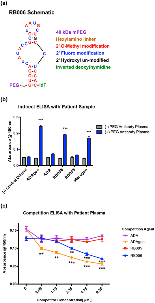
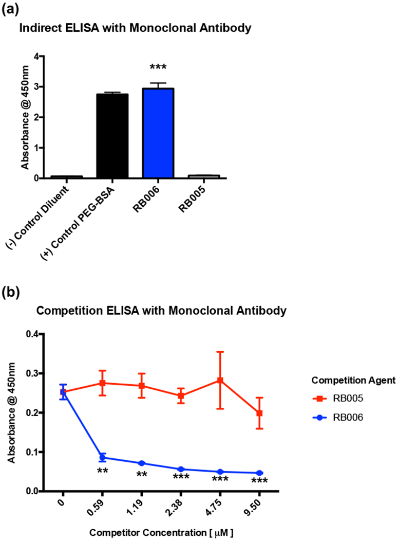
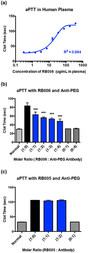
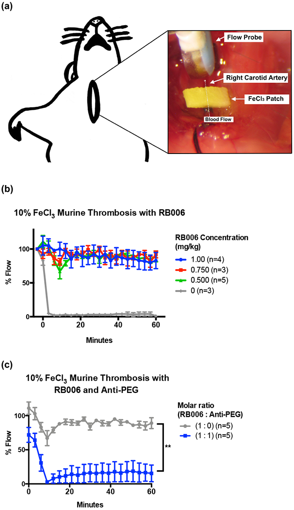
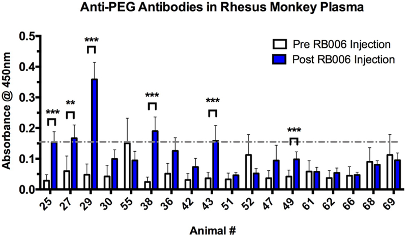
Biopharmaceuticals have become increasingly attractive therapeutic agents and are often PEGylated to enhance their pharmacokinetics and reduce their immunogenicity. However, recent human clinical trials have demonstrated that administration of PEGylated compounds can evoke anti-PEG antibodies. Considering the ubiquity of PEG in commercial products and the presence of pre-existing anti-PEG antibodies in patients in large clinical trials evaluating a PEG-modified aptamer, we investigated how anti-PEG antibodies effect the therapeutic activities of PEGylated RNA aptamers. We demonstrate that anti-PEG antibodies can directly bind to and inhibit anticoagulant aptamer function in vitro and in vivo. Moreover, in parallel studies we detected the presence of anti-PEG antibodies in nonhuman primates after a single administration of a PEGylated aptamer. Our results suggest that anti-PEG antibodies can limit the activity of PEGylated drugs and potentially compromise the activity of otherwise effective therapeutic agents.

Keywords: ELISA (enzyme-linked immunosorbent assay); PEG (polyethylene glycol); PEGylation; RB006; REGULATE-PCI; aPTT (activated partial thromboplastin time); anti-PEG antibodies; aptamer; hypersensitivity; rhesus monkeys.


Conflict of interest statement

Declaration of Interests:

The authors have declared no conflict of interest exists. However, Duke University has submitted patent applications on the anticoagulant aptamers.

Figures

Figure 1-
Figure 1-. PEGylated aptamers are recognized by anti-PEG IgGs in patient plasma
(a) Schematic representation of the PEGylated RNA aptamer RB006. (b) Indirect ELISA demonstrates that plasma from anti-PEG IgG positive patients (blue) exclusively detects PEGylated compounds including the PEGylated RNA aptamers RB006 and Macugen and the PEGylated proteins ADAgen (PEGylated adenosine deaminase). However, patient plasma containing anti-PEG antibodies did not detect the non-PEGylated controls, including a non-PEGylated version of ADAgen, termed ADA, and a non-PEGylated version of RB006, termed RB005. Additionally, patient plasma that is negative for anti-PEG IgGs (grey) did not detect any of the PEGylated compounds. Data represent the mean ± SD of technical triplicates. *** denotes p-value < 0.001, using a t-test against (−) PEG plasma. (c) Competition ELISA demonstrates that patient-derived anti-PEG antibodies bind with more affinity to PEGylated verses unPEGylated compounds, as shown by a decrease in absorbance with increasing concentrations of either the PEGylated aptamer RB006 (blue) or the PEGylated protein ADAgen (orange). No change was seen with the unPEGylated controls RB005 (red) and ADA (purple). Data represents the mean ± SD of technical triplicates. ** denotes p-value<0.01, *** denotes p-value < 0.001, using t-test verses unPEGylated compounds.
Figure 2-
Figure 2-. PEGylated aptamers are recognized by monoclonal anti-PEG IgGs
(a) A commercially available mouse monoclonal anti-PEG antibody specific for PEG recognizes PEGylated aptamer RB006 (blue) and control PEGylated-BSA (black) but does not recognize the unPEGylated aptamer RB005 (grey) through indirect ELISA. Data represent the mean ± SD of technical triplicates. ** denotes p-value <0.01, *** denotes p-value < 0.001, using a t-test against unPEGylated RNA aptamer RB005. (b) Competition ELISA demonstrates that monoclonal anti-PEG IgGs bind to PEGylated aptamer RB006 (blue) with more affinity than to unPEGylated control aptamer RB005 (red). Data represent the mean ± SD of technical triplicates. ** denotes p-value<0.01, *** denotes p-value < 0.001, using a t-test against unPEGylated RNA Aptamer RB005.
Figure 3-
Figure 3-. Aptamer target binding is inhibited in the presence of anti-PEG antibody
Surface plasmon resonance (SPR) was used to test the binding of RB006 to its target protein FIXa both alone and in the presence of anti-PEG IgG. Factor IXa was covalently attached to the chip surface and binding was measured in Response Units (RU) after injection of either RB006 alone (red and green, replicates) or RB006 in the presence of increasing molar ratios of monoclonal anti-PEG antibody (pink and blue respectively). Data represent a single experiment, though the effect was reproducible in two other replicate experiments. (Inset) A decrease in max binding (RU) was observed when aptamer and antibody were premixed and injected over the surface of the chip. The order of listed samples reflects the order of sample injections. The chip surface was uncompromised throughout the duration of the experiment as a similar Rmax was seen with the aptamer only samples injected at the beginning of the assay (red) and at the end of the assay (green).
Figure 4-
Figure 4-. Aptamer function is inhibited in the presence of anti-PEG antibodies in vitro
(a) Activated partial thromboplastin time (aPTT) measurement of the anticoagulant activity of RNA aptamer RB006 (blue) in pooled normal human plasma demonstrates aptamer mediated clotting time extension occurs in a dose dependent manner. A max clotting time of ~125 seconds is observed, which is significantly above the normal clotting time of approximately 30 seconds. Data represent the mean ± SD of technical replicates (N=4 wells). (b) aPTT measurements of human plasma clotting time in the presence of RB006 with or without anti-PEG IgG in the reaction. Decreases in aptamer mediated clot-time extension with increasing concentrations of monoclonal anti-PEG IgG (blue) provide evidence for antibody mediated inhibition of RB006 function. Data represent the mean ± SD of technical replicates (N=6 wells) pooled from triplicate experiments. *** denotes p-value < 0.001, using t-test against aptamer only (1:0) (black). (c) aPTT measurements of human plasma clotting time in the presence of the unPEGylated control aptamer RB005 with or without anti-PEG IgG demonstrates no change in clotting time, suggesting that anti-PEG IgG only inhibits the function of PEGylated aptamer. Data represent the mean ± SD of technical duplicates.
Figure 5-
Figure 5-. Anticoagulant aptamer activity is inhibited in the presence of anti-PEG antibodies in vivo
(a) Schematic represents the murine model of thrombosis used to analyze the antithrombotic / anticoagulant properties of aptamers and anti-PEG antibodies in real time. A ferric chloride (FeCl3) patch is used to induce endothelial damage to the carotid artery, and blood flow through the artery is measured via a distal flow probe. Percent blood flow at various time points post-intravenous (IV) administration of the aptamer was normalized to the blood flow prior to placement of the FeCl3 patch, with time zero indicating the time of patch removal. (b) Increasing concentrations of PEGylated RB006 (green, red, blue) demonstrate the antithrombotic activity compared to buffer control (grey) in the murine model of thrombosis. Data represent the mean ± SEM of technical replicates as described in the figure legend. (c) Addition of anti-PEG IgG at a 1:1 molar ratio to the PEGylated RB006 (blue) diminishes vessel patency in the murine thrombosis model as shown by a decrease in % Flow compared to aptamer alone (grey). For both treatment groups, RB006 was used at a concentration of 0.5 mg/kg. Data represent the mean ± SEM of technical replicates as described. **indicates p-value <0.01, using 2-way ANOVA vs. aptamer alone (1:0).
Figure 6-
Figure 6-. Anti-PEG antibodies can be detected in rhesus plasma after a single administration of RB006
Indirect ELISA was used to measure the level of anti-PEG IgG in plasma before and after RB006 administration, with PEG-BSA coated 96-well plates followed by the addition of plasma for recognition and IgG rhesus monkey specific antibody for detection. Six out of 18 healthy rhesus monkeys tested had significant increases in anti-PEG IgG levels between the pre-aptamer injection samples (white) and post-aptamer injection samples (blue), with four of the animals reaching anti-PEG IgG levels greater than 2 SD from the mean of all pre-injection absorbance values (grey dashed line). The route of administration and dosing of the aptamer as well as the time of sample collection are described in Table 1. The animals selected and their numbers are from the original numerical designation in Figure S1. Data represent the mean ± SEM of technical triplicates. ** indicates p-value <0.01, ***indicates p-value <0.001, using a t-test.

Comment in

References

    1. Aberle JH, Aberle SW, Redlberger-Fritz M, Sandhofer MJ, and Popow-Kraupp T (2010). Human metapneumovirus subgroup changes and seasonality during epidemics. Pediatr Infect Dis J 29, 1016–1018. - PubMed
    1. Abuchowski A, McCoy JR, Palczuk NC, van Es T, and Davis FF (1977a). Effect of covalent attachment of polyethylene glycol on immunogenicity and circulating life of bovine liver catalase. J Biol Chem 252, 3582–3586. - PubMed
    1. Abuchowski A, van Es T, Palczuk NC, and Davis FF (1977b). Alteration of immunological properties of bovine serum albumin by covalent attachment of polyethylene glycol. J Biol Chem 252, 3578–3581. - PubMed
    1. Chan MY, Cohen MG, Dyke CK, Myles SK, Aberle LG, Lin M., Walder J, Steinhubl SR, Gilchrist IC, Kleiman NS, et al. (2008). Phase 1b randomized study of antidote-controlled modulation of factor IXa activity in patients with stable coronary artery disease. Circulation 117, 2865–2874. - PubMed
    1. Cohen MG, Purdy DA, Rossi JS, Grinfeld LR, Myles SK, Aberle LH, Greenbaum AB, Fry E, Chan MY, Tonkens RM, et al. (2010). First clinical application of an actively reversible direct factor IXa inhibitor as an anticoagulation strategy in patients undergoing percutaneous coronary intervention. Circulation 122, 614–622. - PubMed

Publication types

MeSH terms