Review
. 2019 Feb 22:8:F1000 Faculty Rev-211.
doi: 10.12688/f1000research.16234.1. eCollection 2019.

Recent innovations in membrane-protein structural biology

Affiliations
Review

Recent innovations in membrane-protein structural biology

James P Allen. F1000Res. .

Abstract

Innovations are expanding the capabilities of experimental investigations of the structural properties of membrane proteins. Traditionally, three-dimensional structures have been determined by measuring x-ray diffraction using protein crystals with a size of least 100 μm. For membrane proteins, achieving crystals suitable for these measurements has been a significant challenge. The availabilities of micro-focus x-ray beams and the new instrumentation of x-ray free-electron lasers have opened up the possibility of using submicrometer-sized crystals. In addition, advances in cryo-electron microscopy have expanded the use of this technique for studies of protein crystals as well as studies of individual proteins as single particles. Together, these approaches provide unprecedented opportunities for the exploration of structural properties of membrane proteins, including dynamical changes during protein function.

Keywords: X-ray free electron laser; cryo-electron microscopy; protein crystallography; protein dynamics.


Conflict of interest statement

No competing interests were disclosed.No competing interests were disclosed.No competing interests were disclosed.No competing interests were disclosed.

Figures

Figure 1.
Figure 1.. Model of the bacterial reaction center buried within a membrane.
Shown are the protein subunits of the reaction center from Rhodobacter sphaeroides (orange, green, purple subunits with blue cofactors) that are positioned within an artificial membrane (yellow carbon chains with white head groups) based upon a range of spectroscopic studies (modified from 1).
Figure 2.
Figure 2.. Three-dimensional structures of reaction centers from Blastochloris viridis and Rhodobacter sphaeroides
The arrangement of the bacteriochlorophyll and quinone cofactors (red) is shown. The two core protein subunits—L (yellow) and M (cyan)—and the H subunit (green) are all conserved, whereas the tetraheme cytochrome subunit (orange) is found only in reaction centers from B. viridis. (PDB files 1PRC and 4RCR , .)
Figure 3.
Figure 3.. Three-dimensional structure of the human β 2 adrenergic receptor (cyan) showing the presence of seven-transmembrane helices that are conserved among G protein–coupled receptors surrounding the agonist carazolol (red).
For crystallization, a lysozyme domain (wheat) was added, replacing one of the connecting loops (PDB file 2RH1 ).
Figure 4.
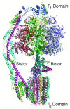
Figure 4.. Three-dimensional structure of ATP synthase.
The backbones of the 14 protein subunits are shown (and each protein subunit is colored differently). During catalysis, the hydrophilic F 1 domain rotates relative to the domain that is embedded in the cell membrane. This rotation modifies the protein environment of the catalytic site, resulting in the production of ATP. The complex can be considered to be a rotary motor with a stator and rotor rotation that is powered by a transfer of protons across the cell membrane (PDB file 6CP6 ).
Figure 5.
Figure 5.. Three-dimensional structures of bacteriorhodopsin at different periods of time following illumination.
Absorption of light by the retinal results in a trans-cis isomerization, which starts the process of proton transfer. Structural changes are evident on very fast timescales. The structures are shown for the retinal and a few surrounding residues, Leu 93, Thr 178, Trp 182, Lys 216, and Phe 219 at the initial time (red), 760 ns after excitation (green), and 1.7 ms after excitation (cyan). (PDB files 5B6V, 5B6X, and 5B6Z .)

References

    1. Kuang L, Olson TL, Lin S, et al. : Interface for Light-Driven Electron Transfer by Photosynthetic Complexes Across Block Copolymer Membranes. J Phys Chem Lett. 2014;5(5):787–91. 10.1021/jz402766y - DOI - PubMed
    1. Thal DM, Glukhova A, Sexton PM, et al. : Structural insights into G-protein-coupled receptor allostery. Nature. 2018;559(7712):45–53. 10.1038/s41586-018-0259-z - DOI - PubMed
    2. F1000 Recommendation

    1. Renaud JP, Chari A, Ciferri C, et al. : Cryo-EM in drug discovery: achievements, limitations and prospects. Nat Rev Drug Discov. 2018;17(7):471–92. 10.1038/nrd.2018.77 - DOI - PubMed
    2. F1000 Recommendation

    1. Henderson R, Unwin PN: Three-dimensional model of purple membrane obtained by electron microscopy. Nature. 1975;257(5521):28–32. 10.1038/257028a0 - DOI - PubMed
    1. Deisenhofer J, Epp O, Miki K, et al. : Structure of the protein subunits in the photosynthetic reaction centre of Rhodopseudomonas viridis at 3Å resolution. Nature. 1985;318(6047):618–24. 10.1038/318618a0 - DOI - PubMed
    2. F1000 Recommendation

Publication types

Substances

LinkOut - more resources