. 2019 Feb 21:7:13.
doi: 10.1186/s40560-019-0368-2. eCollection 2019.

Physician agreement on the diagnosis of sepsis in the intensive care unit: estimation of concordance and analysis of underlying factors in a multicenter cohort

Affiliations

Physician agreement on the diagnosis of sepsis in the intensive care unit: estimation of concordance and analysis of underlying factors in a multicenter cohort

Bert K Lopansri et al. J Intensive Care. .

Abstract

Background: Differentiating sepsis from the systemic inflammatory response syndrome (SIRS) in critical care patients is challenging, especially before serious organ damage is evident, and with variable clinical presentations of patients and variable training and experience of attending physicians. Our objective was to describe and quantify physician agreement in diagnosing SIRS or sepsis in critical care patients as a function of available clinical information, infection site, and hospital setting.

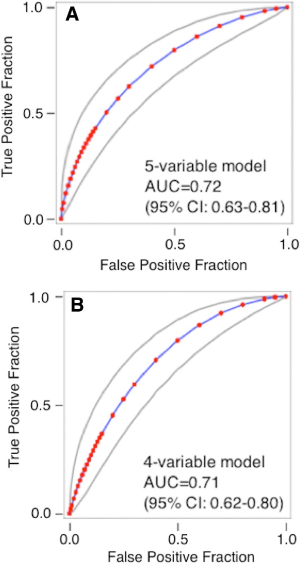
Methods: We conducted a post hoc analysis of previously collected data from a prospective, observational trial (N = 249 subjects) in intensive care units at seven US hospitals, in which physicians at different stages of patient care were asked to make diagnostic calls of either SIRS, sepsis, or indeterminate, based on varying amounts of available clinical information (clinicaltrials.gov identifier: NCT02127502). The overall percent agreement and the free-marginal, inter-observer agreement statistic kappa (κ free) were used to quantify agreement between evaluators (attending physicians, site investigators, external expert panelists). Logistic regression and machine learning techniques were used to search for significant variables that could explain heterogeneity within the indeterminate and SIRS patient subgroups.

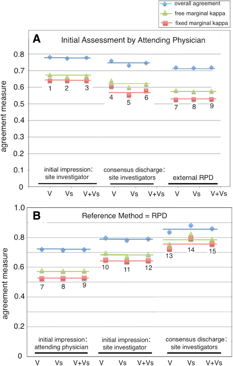
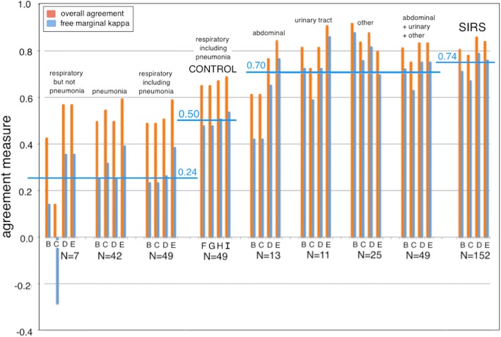
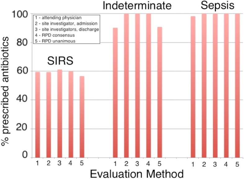
Results: Free-marginal kappa decreased between the initial impression of the attending physician and (1) the initial impression of the site investigator (κ free 0.68), (2) the consensus discharge diagnosis of the site investigators (κ free 0.62), and (3) the consensus diagnosis of the external expert panel (κ free 0.58). In contrast, agreement was greatest between the consensus discharge impression of site investigators and the consensus diagnosis of the external expert panel (κ free 0.79). When stratified by infection site, κ free for agreement between initial and later diagnoses had a mean value + 0.24 (range - 0.29 to + 0.39) for respiratory infections, compared to + 0.70 (range + 0.42 to + 0.88) for abdominal + urinary + other infections. Bioinformatics analysis failed to clearly resolve the indeterminate diagnoses and also failed to explain why 60% of SIRS patients were treated with antibiotics.

Conclusions: Considerable uncertainty surrounds the differential clinical diagnosis of sepsis vs. SIRS, especially before organ damage has become highly evident, and for patients presenting with respiratory clinical signs. Our findings underscore the need to provide physicians with accurate, timely diagnostic information in evaluating possible sepsis.

Keywords: Diagnosis; Intensive care; Inter-observer agreement; Sepsis.


Conflict of interest statement

Ethics approval was gained from the relevant institutional review boards: Intermountain Medical Center/Latter Day Saints Hospital (1024931); Johns Hopkins Hospital (IRB00087839); Rush University Medical Center (15111104-IRB01); Loyola University Medical Center (208291); Northwell Healthcare (16-02-42-03); and Grady Memorial Hospital (000-87806).This manuscript does not contain any individual person’s data in any form. Therefore, consent for publication is not required.The authors have read the journal’s policy and declare the following competing interests: LM, TDY, AR, RBB, RAB, and TS are current or past employees and/or shareholders of Immunexpress.Springer Nature remains neutral with regard to jurisdictional claims in published maps and institutional affiliations.

Figures

Fig. 1
Fig. 1
Agreement between diagnostic methods. a Comparisons 1, 2, and 3: initial assessment by attending physician vs. initial assessment by site investigator. Comparisons 4, 5, and 6: initial assessment by attending physician vs. discharge assessment by site investigators. Comparisons 7, 8, and 9: initial assessment by attending physician vs. external RPD. Agreement with the initial assessment by attending physician decreases (fixed-marginal kappa κfixed 0.64 ➔ 0.58 ➔ 0.53) as more diagnostic information becomes available, as physician training and experience increases, and as time pressure to make a diagnostic call decreases. b Comparisons 7, 8, and 9: initial assessment by attending physician vs. external RPD. Comparisons 10, 11, and 12: initial assessment by site investigator vs. external RPD. Comparisons 13, 14, and 15: consensus discharge assessment by site investigators vs. external RPD. The numerals and symbols in this figure have the following meanings: 1, 4, 7, 10, and 13: VENUS cohort (V; 129 subjects); 2, 5, 8, 11, and 14: VENUS supplemental cohort (Vs; 120 subjects); 3, 6, 9, 12, 15: VENUS + VENUS supplemental cohorts (V + Vs; 249 subjects); blue diamonds = overall agreement; green triangles = free-marginal kappa κfree; red squares = fixed-marginal kappa κfixed
Fig. 2
Fig. 2
Plot of percent overall agreement and free-marginal kappa (κfree) for diagnostic method comparisons stratified by site of infection. For each infection site, the following comparisons were performed: (B) initial assessment by attending physician vs. consensus discharge assessment by site investigators; (C) initial assessment by attending physician vs. external RPD; (D) initial assessment by site investigator vs. consensus discharge assessment; and (E) initial assessment by site investigator vs. external RPD. As a control, the following comparisons were performed for respiratory infection samples including pneumonia (N = 49): (F) consensus discharge assessment vs. external RPD; (G) RPD panelists 1 vs. 2; (H) RPD panelists 1 vs. 3; (I) RPD panelists 2 vs. 3. Note: the number of subjects in the various categories add up to 250 (not 249) because the infection site for one sepsis case was diagnosed as both abdominal and pneumonia. “SIRS” indicates that no site of infection was identified. Horizontal blue bars indicate average values for the free-marginal kappa statistic, over the indicated comparisons
Fig. 3
Fig. 3
Logistic regression analysis to distinguish indeterminates from patients with either sepsis or SIRS. a Logistic regression model for sepsis vs. indeterminates. The model used consensus discharge diagnosis by the site investigators as the comparator and analyzed 64 septic patients and 23 indeterminates. The predictor variable is given by the following equation: y = 0.4249 + 0.3672 * SeptiScore + 0.1232 * WBC.Max − 0.0245 * WBC.Min − 0.0269 * MAP.Max. This equation gives AUC = 0.79 (95% CI 0.68–0.90) in ROC curve analysis. b Logistic regression model for SIRS vs. indeterminates. The model used consensus discharge diagnosis by the site investigators as the comparator and analyzed 73 SIRS patients and 15 indeterminates. The predictor variable is given by the following equation: y = 3.1742–0.2548 * log2 PCT − 0.3913 * SeptiScore. This equation gives AUC = 0.81 (95% CI 0.69–0.92) in ROC curve analysis. Additional file 6 provides further details of the analysis. Abbreviations: AUC, area under curve; MAP.Max, maximum mean arterial blood pressure; PCT, procalcitonin; ROC, receiver operating characteristic curve; WBC.Max, maximum white blood cell count; WBC.Min, minimum white blood cell count
Fig. 4
Fig. 4
Analysis of subjects treated with therapeutic antibiotics as a function of diagnosis, evaluation method, and cohort: fraction of subjects treated. The case report forms indicated whether or not particular patients were treated with therapeutic antibiotics. A diagnosis of SIRS, indeterminate, or sepsis was made by (1) attending physician at admission, (2) site investigator at admission, (3) site investigators’ consensus at discharge, (4) consensus RPD, or (5) unanimous RPD
Fig. 5
Fig. 5
Analysis of subjects treated with therapeutic antibiotics as a function of diagnosis, evaluation method, and cohort: logistic regression models. a Discrimination of SIRS patients who were treated vs. not treated with therapeutic antibiotics, using a five-variable logistic model (y = − 17.8210 − 0.0200 * MAP.Min + 0.0128 * HR.Max + 0.4540 * Temp.Max + 0.0906 * Hospital.LoS + 0.2472 * N.SIRS). The model gave AUC 0.72 (95% CI 0.63–0.81) in ROC curve analysis. b Discrimination of SIRS patients who were treated vs. not treated with therapeutic antibiotics using a four-variable logistic model (y = − 16.5106 - 0.0239 * MAP.Min + 0.0125 * HR.Max + 0.4372 * Temp.Max + 0.2386 * N.SIRS). The model gave AUC 0.71 (95% CI 0.62–0.80) in ROC curve analysis. Additional file 7 provides further details. Abbreviations: AUC, area under curve; Hospital.LoS, length of stay in hospital; HR.Max, maximum heart rate; MAP.Min, minimum mean arterial blood pressure; N.SIRS, number of SIRS criteria met; ROC, receiver operating characteristic curve; Temp.Max, maximum core temperature; WBC.Max, maximum white blood cell count; WBC.Min, minimum white blood cell count

References

    1. Vincent JL. The clinical challenge of sepsis identification and monitoring. PLoS Med. 2016;13:e1002022–10. 10.1371/journal.pmed.1002022. - PMC - PubMed
    1. Phua J, Ngerng W, See K, Tay C, Kiong T, Lim H, et al. Characteristics and outcomes of culture-negative versus culture-positive severe sepsis. Crit Care. 2013;17:R202. 10.1186/cc12896. - PMC - PubMed
    1. Dunne WM Jr. Laboratory diagnosis of sepsis? No SIRS, not just yet. J Clin Microbiol. 2015;53:2404–9. 10.1128/JCM.03681-14. - PMC - PubMed
    1. Dellinger RP, Levy MM, Rhodes A, Annane D, Gerlach H, Opal SM, et al. Surviving Sepsis Campaign: international guidelines for management of severe sepsis and septic shock, 2012. Intensive Care Med. 2013;39:165–228. 10.1007/s00134-012-2769-8. - PMC - PubMed
    1. Bai X, Yu W, Ji W, Lin Z, Tan S, Duan K, et al. Early versus delayed administration of norepinephrine in patients with septic shock. Crit Care. 2014;18:532. 10.1186/s13054-014-0532-y. - PMC - PubMed

Associated data