. 2019;7(2):81-90.
doi: 10.1080/21678707.2019.1562334. Epub 2019 Jan 9.

Progress in understanding Friedreich's ataxia using human induced pluripotent stem cells

Affiliations

Progress in understanding Friedreich's ataxia using human induced pluripotent stem cells

Anna M Schreiber et al. Expert Opin Orphan Drugs. 2019.

Abstract

Introduction: Friedreich's ataxia (FRDA) is an autosomal recessive multisystem disease mainly affecting the peripheral and central nervous systems, and heart. FRDA is caused by a GAA repeat expansion in the first intron of the frataxin (FXN) gene, that leads to reduced expression of FXN mRNA and frataxin protein. Neuronal and cardiac cells are primary targets of frataxin deficiency and generating models via differentiation of induced pluripotent stem cells (iPSCs) into these cell types is essential for progress towards developing therapies for FRDA.

Areas covered: This review is focused on modeling FRDA using human iPSCs and various iPSC-differentiated cell types. We emphasized the importance of patient and corrected isogenic cell line pairs to minimize effects caused by biological variability between individuals.

Expert opinion: The versatility of iPSC-derived cellular models of FRDA is advantageous for developing new therapeutic strategies, and rigorous testing in such models will be critical for approval of the first treatment for FRDA. Creating a well-characterized and diverse set of iPSC lines, including appropriate isogenic controls, will facilitate achieving this goal. Also, improvement of differentiation protocols, especially towards proprioceptive sensory neurons and organoid generation, is necessary to utilize the full potential of iPSC technology in the drug discovery process.

Keywords: Friedreich’s ataxia; GAA repeat expansion; differentiation; frataxin; induced pluripotent stem cells.


Conflict of interest statement

Declaration of interest M Napierala is in part supported by CRISPRx Therapeutics Inc. The sponsor had no role or any influence of the content of this manuscript. The authors have no other relevant affiliations or financial involvement with any organization or entity with a financial interest in or financial conflict with the subject matter or materials discussed in the manuscript.

Figures

Figure 1.
Figure 1.
Generation of human iPSCs and further differentiation schemes to create FRDA affected cell types. The iPSCs can be obtained from different somatic cell types including blood cells, keratinocytes, and fibroblasts. In addition to the classic “Yamanaka” transcription factors (Oct3/4, Sox2, c-Myc and Klf4), many variants of transcription factors have been reported to be both sufficient and necessary to increase the efficiency of the reprogramming process, including additional auxiliary factors or small molecules. Only cell types that are the most relevant to FRDA pathology are depicted.
Figure 2.
Figure 2.
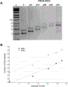
Continuous expansions observed in FRDA iPSCs. A. The FRDA fibroblast line GM04078 (Coriell Cell Repositories) harboring ~ 340/450 GAA repeats (GAA1/GAA2 alleles, lane F) was reprogrammed to iPSCs using retroviral transduction of pluripotency transcription factors. Somatic reprogramming was associated with expansion to 410/540 GAAs in passage 0 of the iPSC culture (p0). Subsequent passaging resulted in continuous expansions reaching ~700/860 GAAs after passage 27 (p27). Approximately 200 days of culture (reprogramming followed by 27 passages of the iPSCs) was sufficient to double the number of GAAs observed in the parental fibroblasts. B. Expansion of the GAA repeats is observed for both GAA1 and GAA2 alleles. An increase of 10 – 11 GAAs per passage was detected for each allele.
Figure 3.
Figure 3.
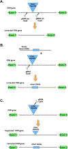
Use of genome editing to generate isogenic pairs of FRDA and corrected iPSC lines. For simplicity correction of a single allele harboring the expanded repeats is shown. Expanded GAAs are depicted as blue triangles while short, nonpathogenic GAAs appear as blue rectangles. Only exons 1 and 2 and intron 1 of FXN are shown. Also, only the Cas9/gRNA system is depicted as the DNA DSB or nick inducer. A. Excision of the expanded GAAs using a pair of nucleases cleaving upstream and downstream of the GAAs. A fragment of intron 1 is removed together with the entire GAA tract. B. Replacement of the expanded GAAs with a homologous intron 1 DNA fragment containing short GAAs via HDR. No or minimal changes to the nucleotide composition of intron 1 are introduced. C. Generation of GAA tract contractions using a repeat targeting Cas9 nickase. Several different products can be expected, which contain shorter pathogenic GAA tracts or a stretch of the repeats within the unaffected range. Two distinct possible products of editing are depicted: “improved” and corrected.

Similar articles

Cited by

References

    1. Koeppen AH, Friedreich’s ataxia: pathology, pathogenesis, and molecular genetics. J Neurol Sci 2011;303(1–2):1–12. - PMC - PubMed
    1. Delatycki MBLA Corben, Clinical features of Friedreich ataxia. J Child Neurol 2012;27(9):1133–7. - PMC - PubMed
    1. Campuzano V, Montermini L, Molto MD, et al., Friedreich’s ataxia: autosomal recessive disease caused by an intronic GAA triplet repeat expansion. Science 1996;271(5254):1423–7. - PubMed
    1. Durr A, Cossee M, Agid Y, et al., Clinical and genetic abnormalities in patients with Friedreich’s ataxia. N Engl J Med 1996;335(16):1169–75. - PubMed
    1. Clark E, Butler JS, Isaacs CJ, et al., Selected missense mutations impair frataxin processing in Friedreich ataxia. Ann Clin Transl Neurol 2017;4(8):575–584. - PMC - PubMed

LinkOut - more resources