

Benign proliferative epithelial lesions of oral mucosa are infrequently associated with α-, β-, or γ human papillomaviruses
- PMID: 30828618
- PMCID: PMC6383307
- DOI: 10.1002/lio2.222
Benign proliferative epithelial lesions of oral mucosa are infrequently associated with α-, β-, or γ human papillomaviruses
Abstract
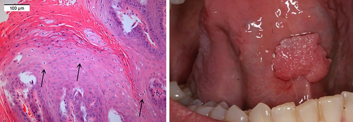
Background: Oral papillomas and verruca vulgaris have been associated with human papillomavirus (HPV) infection. However, approximately half of these have remained HPV-negative when tested for mucosal HPV genotypes. In this study, we evaluated presence of α-, β-, and γ-HPVs in benign papillary and verrucous lesions.
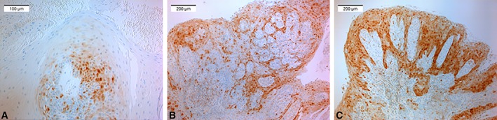
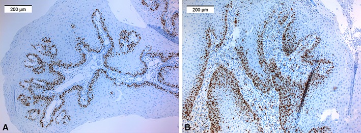
Methods: Eighty-three clinical lesions with suspected HPV etiology were analyzed for HPV types of genus α (n = 24), β (n = 46), and γ (n = 52). Immunohistochemistry was used for p16 as a possible surrogate marker of high-risk HPV, accompanied by Ki-67 proliferation marker.
Results: Altogether, α-HPVs were detected in 6.4%, β-HPVs in 2.4%, and γ-HPV in 4.8%. The following genotypes were identified: HPV6, 8, 11, 16, 22, 161, and 170. Neither Ki-67 nor p16 positivity alone were associated with HPV but combined staining showed significant inverse association (P = .042).
Conclusion: HPV infection is found only in a minority of benign verrucous and papillary oral lesions, with the predominance of α-HPVs.
Level of evidence: 4.
Keywords: HPV; Oral; benign; p16; papilloma.
Figures
Similar articles
-
Biology and pathological associations of the human papillomaviruses: a review.Malays J Pathol. 1998 Jun;20(1):1-10. Malays J Pathol. 1998. PMID: 10879257 Review.
-
HPV-Related Papillary Lesions of the Oral Mucosa: A Review.Head Neck Pathol. 2019 Mar;13(1):80-90. doi: 10.1007/s12105-019-01003-7. Epub 2019 Jan 29. Head Neck Pathol. 2019. PMID: 30693456 Free PMC article. Review.
-
P16(INK4A) immunostaining is a strong indicator for high-risk-HPV-associated oropharyngeal carcinomas and dysplasias, but is unreliable to predict low-risk-HPV-infection in head and neck papillomas and laryngeal dysplasias.Int J Cancer. 2014 May 1;134(9):2108-17. doi: 10.1002/ijc.28534. Epub 2013 Oct 21. Int J Cancer. 2014. PMID: 24127203
-
Human Papillomavirus and Risk of Head and Neck Squamous Cell Carcinoma in Iran.Microbiol Spectr. 2022 Aug 31;10(4):e0011722. doi: 10.1128/spectrum.00117-22. Epub 2022 Jun 16. Microbiol Spectr. 2022. PMID: 35708339 Free PMC article.
-
Prevalence of mucosal and cutaneous human papillomaviruses in different histologic subtypes of vulvar carcinoma.Mod Pathol. 2008 Mar;21(3):334-44. doi: 10.1038/modpathol.3801009. Epub 2008 Jan 11. Mod Pathol. 2008. PMID: 18192968
Cited by
-
Role of human papillomavirus infection in the etiology of vulvar cancer in Italian women.Infect Agent Cancer. 2020 Apr 1;15:20. doi: 10.1186/s13027-020-00286-8. eCollection 2020. Infect Agent Cancer. 2020. PMID: 32266002 Free PMC article.
-
Oral Papillomatosis: Its Relation with Human Papilloma Virus Infection and Local Immunity-An Update.Medicina (Kaunas). 2022 Aug 15;58(8):1103. doi: 10.3390/medicina58081103. Medicina (Kaunas). 2022. PMID: 36013570 Free PMC article. Review.
-
Analysis of the anatomical distribution of HPV genotypes in head and neck squamous papillomas.PLoS One. 2023 Aug 11;18(8):e0290004. doi: 10.1371/journal.pone.0290004. eCollection 2023. PLoS One. 2023. PMID: 37566623 Free PMC article.
-
Gingiva squamous-cell carcinoma in a non-smoking patient with occupational exposure to solvent siphoning using mouth: case report and literature review.Front Public Health. 2024 May 2;12:1370767. doi: 10.3389/fpubh.2024.1370767. eCollection 2024. Front Public Health. 2024. PMID: 38756897 Free PMC article. Review.
-
HPV-Associated Benign Squamous Cell Papillomas in the Upper Aero-Digestive Tract and Their Malignant Potential.Viruses. 2021 Aug 17;13(8):1624. doi: 10.3390/v13081624. Viruses. 2021. PMID: 34452488 Free PMC article. Review.
References
-
- Jones HJ. Non‐odontogenic tumours in children. Br Dent J 1965:16:439–447. - PubMed
-
- Knapp MJ. Oral disease in 181,338 consecutive oral examinations. J Am Dent Assoc 1971;83:1288‐1293. - PubMed
-
- Bouquot JE, Gundlach KK. Oral exophytic lesions in 23,616 white Americans over 35 years of age. Oral Surg Oral Med Oral Pathol 1986;62:284–291. - PubMed
-
- Axell T. A prevalence study of oral mucosal lesions in an adult Swedish population. Odontol Revy Suppl 1976;36:1–103. - PubMed
-
- Abbey LM, Page DG, Sawyer DR. The clinical and histopathologic features of a series of 464 oral squamous cell papillomas. Oral Surg Oral Med Oral Pathol 1980:49:419–428. - PubMed
Grants and funding
LinkOut - more resources
Full Text Sources
Research Materials