Review
. 2019 Jan 10;4(1):181-187.
doi: 10.1002/lio2.238. eCollection 2019 Feb.

Palatal anatomy for sleep apnea surgery

Affiliations
Review

Palatal anatomy for sleep apnea surgery

Ewa Olszewska et al. Laryngoscope Investig Otolaryngol. .

Abstract

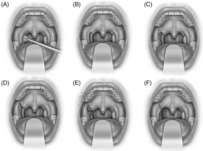
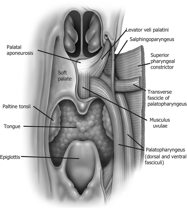
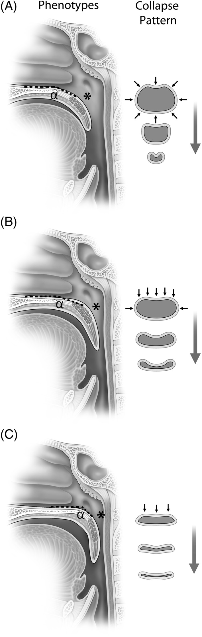
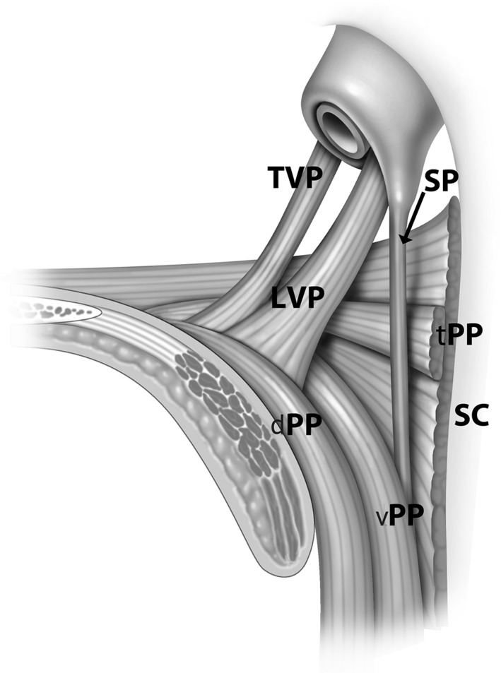
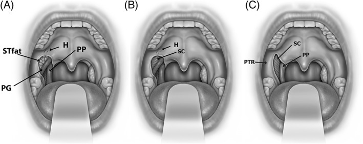
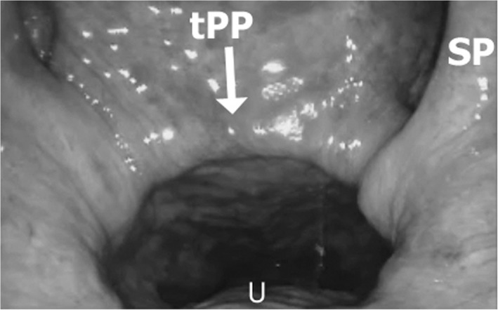
The goal of this review is to advance the understanding of the muscular and soft tissue palatal anatomy as it relates to palatal surgery for sleep apnea and the phenotypic variations that generate the shape and collapsibility of the retropalatal airway. Anatomically, the soft palate has both a proximal and distal segments separated by the palatal genu. The proximal palatal segment has a variable angle from the hard palate (ie, alpha angle) determined by the position and length of the levator veli palatini muscle. The palatopharyngeus muscle (PP) is a major defining element of the palate and lateral pharyngeal wall and forms the medial wall of the lateral palatal space. It is composed of two divisions: the longitudinal palatopharyngeus fasciculi which acts to elevate the pharynx and depress the soft palate and the transverse palatopharyngeus fascicle (Passavant's ridge) which function is a nasopharyngeal sphincter. The lateral palatal space incorporates the supra-tonsilar fat, and is bounded by muscles that determine the structure of the palate and associated lateral pharyngeal walls. Understanding of palatal muscles and pharyngeal airway phenotypes provides insight into the steps and mechanisms of pharyngoplasty procedures.

Level of evidence: N/A.

Keywords: Palatopharyngeus; anatomy; lateral palatal space; obstructive sleep apnea; palate; pharyngoplasty; phenotypes.


Figures

Figure 1
Figure 1
Expansion sphincterplasty is shown (A) tonsillectomy, (B) exposure of palatopharyngeus and superior constrictor muscles within the tonsillar fossa, (C) lateral palatal incision over lateral palatal space with exposure of supratonsillar fat, (D) removal of fat and fibers of palatoglossus provides exposure to superior constrictor and arching fibers of palatopharyngeus muscles, (E) palatopharyngeus muscle incised 1.5 cm. inferior to the fulcrum of rotation and the pedicle is sutured to pterygomandibular raphe or fibrous tissue lateral to the hamulus, and (F) mucosal closure.
Figure 2
Figure 2
Depiction of the major muscular slings that make up the palate and associated airway.
Figure 3
Figure 3
Palatal phenotypes and pattern of collapse are shown. (A) Oblique pattern demonstrates a less acute alpha (α) angle with airway size larger in an anterior posterior dimension at the hard palate, genu (*), and velum; shape is more circular and collapse pattern is circular. (B) Intermediate pattern demonstrates a less acute alpha angle (α) but narrowing at the genu (*) and velum. (C) Vertical pattern has a more acute alpha (α) angle with narrowing of the airway at the genu (*) and velum; airway shape is coronal and collapse patter is flat (anterior posterior).
Figure 4
Figure 4
Muscles of the palate are shown tensor veli palatini (TVP), levator veli palatini (LVP), salphingopharyngeus (SP), superior pharyngeal constrictor (SC), and transverse fascicle (tPP), dorsal fascicle (dPP), and ventral fascicle (vPP) of the palatopharyngeus muscle (PP).
Figure 5
Figure 5
Anatomy of the lateral palatal space is shown. Exposure of palatoglossus muscle and supratonsillar fat (STfat) (in A), removal of supratonsillar fat in pterygoid humulus area (H) (in B) and palatoglossus muscle (PG) (in C) demonstrate the palatal anatomic relationships of the superior pharyngeal constrictor muscle (SC), palatopharyngeus muscle (PP), and pterygomandibular raphe (PTR).
Figure 6
Figure 6
CT scan imaging showing aberrant internal carotid artery (white arrows).
Figure 7
Figure 7
Transverse fascicle of the palatopharyngeus muscle (tPP) is shown on endoscopic view of the nasopharynx. Salphingopharygeus muscle (SP) courses medially and the tPP inserts into the palate in the soft palate lateral to the uvular muscle (U).

References

    1. Pang KP, Woodson BT. Expansion sphincter pharyngoplasty: a new technique for the treatment of obstructive sleep apnea. Otolaryngol Head Neck Surg 2007;137(1):110–114. - PubMed
    1. Cahali MB. Lateral pharyngoplasty: a new treatment for OSAHS. Laryngoscope 2003;113:1961–1968. - PubMed
    1. MacKay SG, Carney AS, Woods C, et al. Modified uvulopalatopharyngoplasty and coblation channeling of a tongue for obstructive sleep apnea: a multi‐center Australian trial. J Clin Sleep Med 2013;9(2):117–124. - PMC - PubMed
    1. Han D, Ye J, Lin Z, Wang J, Wang J, Zhang Y. Revised uvulopalatopharyngoplasty with uvula preservation and its clinical study. ORL J Otorhinolaryngol Relat Spec 2005;67(4):213–219. - PubMed
    1. Li HY, Lee LA, Kezirian EJ, Nakayama M. Suspension palatoplasty for obstructive sleep apnea—a preliminary study. Sci Rep 2018;8(1):4224. - PMC - PubMed