Review
. 2019 Sep 6;21(9):1118-1130.
doi: 10.1093/neuonc/noz048.

Treatment-induced brain tissue necrosis: a clinical challenge in neuro-oncology

Affiliations
Review

Treatment-induced brain tissue necrosis: a clinical challenge in neuro-oncology

Sebastian F Winter et al. Neuro Oncol. .

Abstract

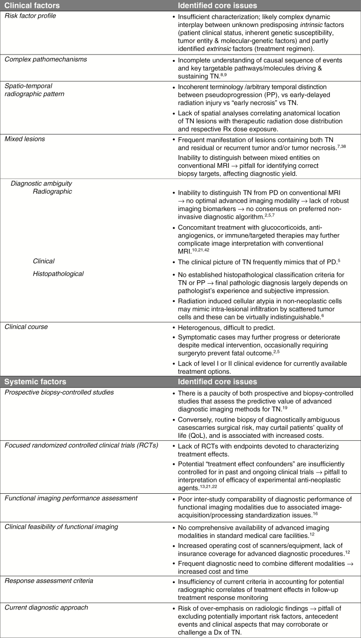
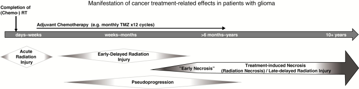
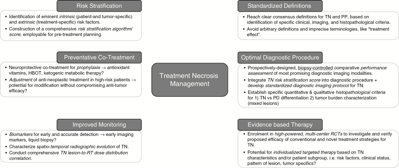
Cancer therapy-induced adverse effects on the brain are a major challenge in neuro-oncology. Brain tissue necrosis (treatment necrosis [TN]) as a consequence of brain directed cancer therapy remains an insufficiently characterized condition with diagnostic and therapeutic difficulties and is frequently associated with significant patient morbidity. A better understanding of the underlying mechanisms, improvement of diagnostic tools, development of preventive strategies, and implementation of evidence-based therapeutic practices are pivotal to improve patient management. In this comprehensive review, we address existing challenges associated with current TN-related clinical and research practices and highlight unanswered questions and areas in need of further research with the ultimate goal to improve management of patients affected by this important neuro-oncological condition.

Keywords: complications; malignant glioma; radiation necrosis; treatment effects; treatment necrosis.


Figures

Fig. 1
Fig. 1
Overview of clinical and systemic factors challenging the study and better understanding of TN. Dx = diagnosis; QoL = quality of life; Rx = radiation.
Fig. 2
Fig. 2
Progressive treatment necrosis (A–C; T1-weighted gadolinium-enhanced axial MRI sequences). (A) A 35-year-old male with right frontal low-grade astrocytoma (World Health Organization grade II) underwent surgical resection followed by TMZ-based chemo-RT treatment. Eight months post-RT completion he developed headaches of increased frequency and was found to have a new nodular focus of enhancement in the right frontal lobe subjacent to the resection cavity, with periventricular and corpus callosum involvement, a biopsy of which revealed TN. (B) Sequential TMZ was resumed and completed over the next 6 months; however, the patient experienced worsening of his symptoms as the region of enhancement continued to expand. (C) Despite initiation of corticosteroid and bevacizumab treatment, he developed progressive left-sided hemiparesis and cognitive decline over the following 2 years, prompting a second biopsy of the continually enhancing lesion, which again confirmed TN. Therapeutic management of symptomatic TN was continued; however, the patient deteriorated further, necessitating a transfer to hospice care, where he eventually passed away 2 years after the second biopsy.
Fig. 3
Fig. 3
Timeline schematic illustrating the temporal manifestation pattern and clinical course of cancer treatment–related effects. Acute and early-delayed types of radiation injury represent transient, reversible neurotoxic phenomena observed within days to weeks, and weeks to several months following chemo-RT. By contrast, TN typically constitutes a late-delayed type of radiation injury observed >6 months post-RT with a frequently irreversible and progressive course; however, concurrent TMZ-based chemo-RT may contribute to increasing incidences of “early necrosis.” Pseudoprogression (PP) likely represents a unique, transient, predominantly radiographic phenomenon encountered in patients with high-grade glioma within the first 3 months of combined TMZ-based chemo-RT. Differentiation between these entities remains a clinical challenge.
Fig. 4
Fig. 4
Schematic illustrating 6 eminent, interdependent research pillars paramount to mapping the field of treatment necrosis management in neuro-oncology. Key research topics and unanswered questions are highlighted.

References

    1. Dietrich J, Winter SF, Klein JP. Neuroimaging of brain tumors: pseudoprogression, pseudoresponse, and delayed effects of chemotherapy and radiation. Semin Neurol. 2017;37(5):589–596. - PubMed
    1. Dietrich J, Klein JP. Imaging of cancer therapy-induced central nervous system toxicity. Neurol Clin. 2014;32(1):147–157. - PubMed
    1. Giglio P, Gilbert MR. Cerebral radiation necrosis. Neurologist. 2003;9(4):180–188. - PubMed
    1. Na A, Haghigi N, Drummond KJ. Cerebral radiation necrosis. Asia Pac J Clin Oncol. 2014;10(1):11–21. - PubMed
    1. Eisele SC, Dietrich J. Cerebral radiation necrosis: diagnostic challenge and clinical management. Rev Neurol. 2015;61(5):225–232. - PubMed

Publication types

MeSH terms

Substances