. 2019 Mar 26;13(3):2913-2926.
doi: 10.1021/acsnano.8b06998. Epub 2019 Mar 4.

Nanoneedle-Mediated Stimulation of Cell Mechanotransduction Machinery

Affiliations

Nanoneedle-Mediated Stimulation of Cell Mechanotransduction Machinery

Catherine S Hansel et al. ACS Nano. .

Abstract

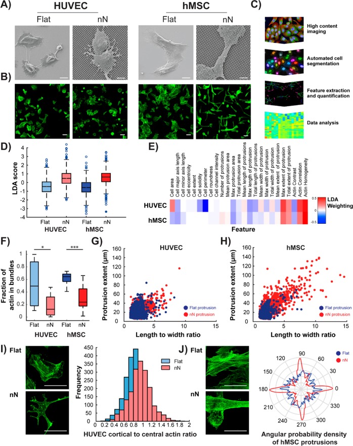
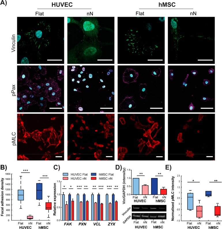
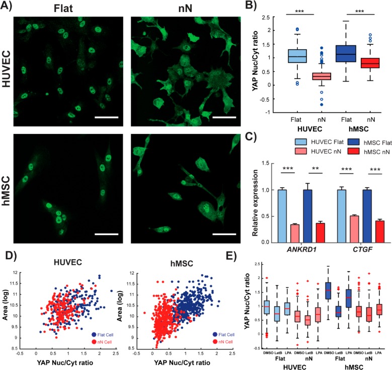
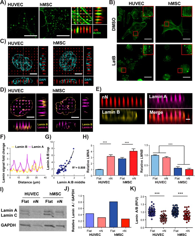
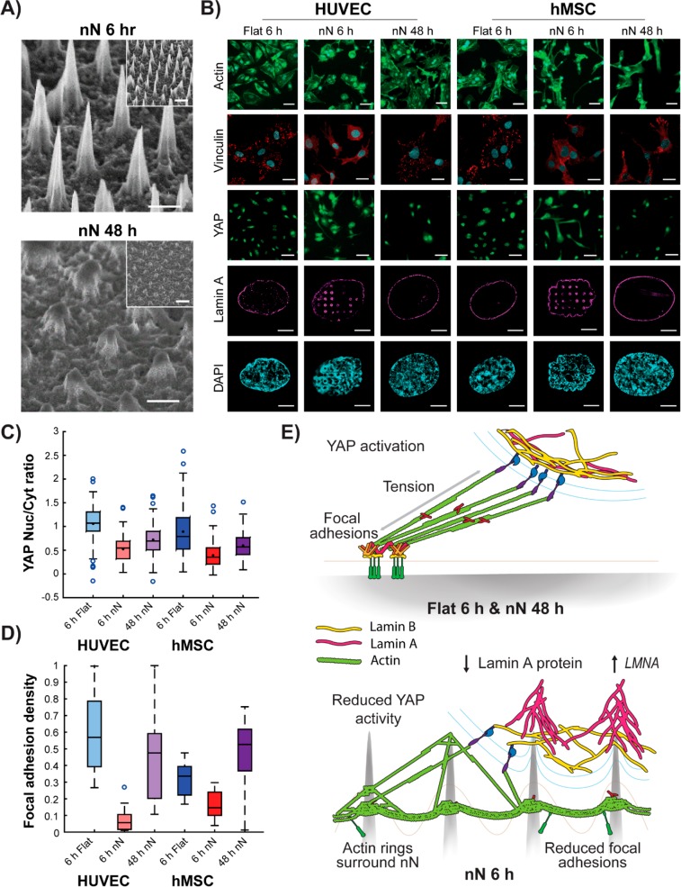
Biomaterial substrates can be engineered to present topographical signals to cells which, through interactions between the material and active components of the cell membrane, regulate key cellular processes and guide cell fate decisions. However, targeting mechanoresponsive elements that reside within the intracellular domain is a concept that has only recently emerged. Here, we show that mesoporous silicon nanoneedle arrays interact simultaneously with the cell membrane, cytoskeleton, and nucleus of primary human cells, generating distinct responses at each of these cellular compartments. Specifically, nanoneedles inhibit focal adhesion maturation at the membrane, reduce tension in the cytoskeleton, and lead to remodeling of the nuclear envelope at sites of impingement. The combined changes in actin cytoskeleton assembly, expression and segregation of the nuclear lamina, and localization of Yes-associated protein (YAP) correlate differently from what is canonically observed upon stimulation at the cell membrane, revealing that biophysical cues directed to the intracellular space can generate heretofore unobserved mechanosensory responses. These findings highlight the ability of nanoneedles to study and direct the phenotype of large cell populations simultaneously, through biophysical interactions with multiple mechanoresponsive components.

Keywords: cell−material interactions; mechanotransduction; nanoneedles; nuclear mechanics; porous silicon; super-resolution microscopy.


Conflict of interest statement

The authors declare no competing financial interest.

Figures

Figure 1
Figure 1
Nanoneedle interaction with HUVECs and hMSCs reduces actin bundling and enhances actin-rich protrusions. (A) SEM images show direct interaction between cells and nN 6 h postseeding. Scale bars = 10 μm. (B) Wide-field immunofluorescence images of the actin cytoskeleton show drastic alterations to cell morphology on nN as compared to that on flat controls (green: phalloidin). HUVECs display a stellate morphology on nN, whereas hMSCs elongate along the nN array. Scale bars = 50 μm. (C) Workflow for extraction, quantification, and analysis of morphometric features using high-content imaging and automated cell segmentation algorithms. (D) Twenty-five features are compared by linear discriminant analysis (LDA) for the two cell types on the flat and nN substrates, and (E) most heavily influenced parameter measured is actin homogeneity. (F) Specific analysis of actin features reveals reduced stress fiber formation (actin bundling) on nN, compared to that on flat substrates for both cell types (box plots, minimum/maximum). (G,H) Image analysis quantification of actin features reveals longer protrusions with high aspect ratios on nN. (I) HUVECs exhibit increased levels of cortical versus central actin on nN (green: phalloidin). (J) hMSC actin cytoskeleton aligns to the nN array lattice. (I,J) Deconvolved maximum projection confocal immunofluorescence images (green: phalloidin). Scale bars = 50 μm. N ≥ 3 experimental replicates for all data; *p < 0.05, ***p < 0.001 between indicated groups.
Figure 2
Figure 2
Nanoneedles inhibit focal adhesion formation and generation of intracellular tension. (A) Confocal maximum projection images 6 h postseeding. On flat substrates, dense vinculin staining is observed in stable focal adhesion (FA) complexes. Strong phosphorylated paxillin (pPax) and phosphorylated myosin light chain (pMLC) signal on flat substrates indicate FA maturation and active actomyosin contractile machinery, respectively. Cells on nN display diffuse vinculin staining and severely reduced pPax and pMLC signal. Scale bars: vinculin = 25 μm; pPax and pMLC = 50 μm. (B) Significant reduction in vinculin signal reveals reduced FA density on nN (box plots, minimum/maximum; N = 3). (C) qPCR indicates that culture on nN yields downregulation in gene expression for multiple FA components (focal adhesion kinase (FAK), paxillin (PAX), vinculin (VCL), and zyxin (ZYX); qPCR, N = 3, mean ± SD). (D) Western blot shows downregulation of vinculin protein expression on nN (HUVEC: N = 2, hMSC: N = 3, mean ± SD). (E) Quantification of pMLC signal intensity via image analysis confirms a significant reduction for both cell types cultured on nN, as compared to their respective controls (box plots, minimum/maximum, N ≥ 4); *p < 0.05, **p < 0.01, ***p < 0.001 between groups as indicated by the lines.
Figure 3
Figure 3
Nanoneedles reduce YAP activity and lessen the correlation between YAP activation and cell spreading. (A) Confocal microscopy shows nuclear YAP protein localization on flat substrates and cytosolic localization on nN (green: YAP). Scale bars = 50 μm. (B) Image analysis quantification of YAP localization shows significant reduction in the nuclear to cytoplasmic ratio of YAP on nN (minimum/maximum; N = 4). (C) qPCR analysis indicates reduced expression of the YAP target genes ankyrin repeat domain 1 (ANKRD1) and connective tissue growth factor (CTGF) (N = 4, mean ± SD). (D) Cell spread area and YAP nuclear localization correlate tightly on flat substrates, but correlation is weakened on nN (N = 3). (E) YAP localization following cell treatment with either the actin depolymerizing agent, LatB, or a small molecule to stimulate actin bundling, LPA. LatB treatment on flat substrates reduces nuclear YAP localization to levels comparable to untreated cells on nN. LatB treatment of cells on nN yields a small reduction in nuclear localization. LPA treatment on flat substrates did not affect YAP localization for HUVECs and marginally decreased this metric for hMSCs. On nN substrates, LPA had little effect on YAP localization. (minimum/maximum; N = 3); *p < 0.05, **p < 0.01, ***p < 0.001 between groups as indicated by the lines.
Figure 4
Figure 4
Nanoneedles interact with mechanoresponsive organelles. (A) Polymerized actin rings form at sites of nN interaction with both HUVECs and hMSCs, (HUVEC: structured illumination microscopy (SIM), single plane; hMSC: deconvolved confocal microscopy, single plane). Green: phalloidin, scale bars = 10 μm. Actin rings were located around the nN (deconvolved confocal z-stack; green: phalloidin, red: nN). Scale bar = 1 μm. (B) Confocal images demonstrate that actin rings still form even when cells are treated with the actin depolymerizing agent, LatB, for the entire 6 h culture period. (Green: phalloidin, single plane). Scale bars = 25 μm. (C) SIM of DAPI-stained nuclei and fluorescent nN shows physical displacement of the nucleus at nN sites (single plane; cyan: DAPI, red: nN). Scale bars = 5 μm. (D) SIM imaging shows lamin B distributing at the base of the nN, with lamin A localising throughout the needle length. (single plane; magenta: lamin A, yellow: lamin B). Scale bars = 5 μm. (E) Reslice images of the xz plane from confocal z-stack images; lamin A signal increases around nN, whereas lamin B remains constant (red: nN, yellow: lamin B, magenta: lamin A). Scale bar = 2 μm. (F) Analysis of fold-change values of lamin A and B intensity along the lower nuclear envelope in resliced confocal images normalized to the signal measured at non-nN locations. (G) Normalized intensity values of lamin A and lamin B at the middle and top of nN. At sites of nuclear remodeling (i.e. lamin A/lamin B > 1) the A/B ratio increases exponentially along the nN axis (R2 = 0.808, n = 69 nN sites, n = 14 cells, N = 3). (H) qPCR analysis of nuclear lamina components shows increased LMNA but not LMNB expression on nN after 6 h in culture. (N = 4, mean ± SD). (I) Western blot and (J) analysis relative to GAPDH control reveal a decrease in protein-level lamin A after 6 h in culture. (K) Quantification of signal intensity for lamin A relative to lamin B images further confirm that a reduction in lamin A occurs following culture on nN substrates (N = 3 experiments); ***p < 0.001 between groups as indicated by the lines.
Figure 5
Figure 5
Nanoneedle degradation recovers mechanoresponsive cell behaviors. (A) SEM images show nN degradation after 48 h in culture. Scale bars = 1 μm, 2 μm inset. (B) Cell phenotype is restored on degraded nN as compared to flat control substrates at 6 h. Cells exhibit a spread actin cytoskeleton (green: phalloidin, scale bars = 50 μm), dense staining of vinculin-rich focal adhesions (red: vinculin, cyan: DAPI, scale bars = 25 μm), nuclear localization of YAP (green, scale bars = 50 μm), and an unimpinged nucleus (magenta: lamin A, cyan: DAPI, scale bars = 5 μm). (C) Image analysis shows a partial return of YAP localization to the nucleus and (D) increased focal adhesion (vinculin) density (box plots, minimum/maximum). (E) Schematic representation of the cell−nN interaction. Cells on flat substrates display firm focal adhesions, which allow for generation of intracellular tension, yielding YAP nuclear localization and subsequent transcriptional activity, and a uniform nuclear lamina composition. nN interfacing limits focal adhesion formation and maturation, directly stimulates actin ring formation, and results in segregation of lamin A and B at the nucleus. Furthermore, lamin A is downregulated at the protein level but upregulated at the gene level in response to interactions with nN.

References

    1. Crowder S. W.; Leonardo V.; Whittaker T.; Papathanasiou P.; Stevens M. M. Material Cues as Potent Regulators of Epigenetics and Stem Cell Function. Cell Stem Cell 2016, 18, 39–52. 10.1016/j.stem.2015.12.012. - DOI - PMC - PubMed
    1. Dalby M. J.; Biggs M. J. P.; Gadegaard N.; Kalna G.; Wilkinson C. D. W.; Curtis A. S. G. Nanotopographical Stimulation of Mechanotransduction and Changes in Interphase Centromere Positioning. J. Cell. Biochem. 2007, 100, 326–338. 10.1002/jcb.21058. - DOI - PubMed
    1. Engler A. J.; Sen S.; Sweeney H. L.; Discher D. E. Matrix Elasticity Directs Stem Cell Lineage Specification. Cell 2006, 126, 677–689. 10.1016/j.cell.2006.06.044. - DOI - PubMed
    1. McMurray R. J.; Gadegaard N.; Tsimbouri P. M.; Burgess K. V.; McNamara L. E.; Tare R.; Murawski K.; Kingham E.; Oreffo R. O. C.; Dalby M. J. Nanoscale Surfaces for the Long-Term Maintenance of Mesenchymal Stem Cell Phenotype and Multipotency. Nat. Mater. 2011, 10, 637–644. 10.1038/nmat3058. - DOI - PubMed
    1. Swift J.; Ivanovska I. L.; Buxboim A.; Harada T.; Dingal P. C. D. P.; Pinter J.; Pajerowski J. D.; Spinler K. R.; Shin J. W.; Tewari M.; Rehfeldt F.; Speicher D. W.; Discher D. E. Nuclear Lamin-A Scales with Tissue Stiffness and Enhances Matrix-Directed Differentiation. Science 2013, 341, 1240104.10.1126/science.1240104. - DOI - PMC - PubMed

Publication types