. 2019 Sep;103(9):1809-1820.
doi: 10.1097/TP.0000000000002683.

Antibody-suppressor CD8+ T Cells Require CXCR5

Affiliations

Antibody-suppressor CD8+ T Cells Require CXCR5

Jason M Zimmerer et al. Transplantation. 2019 Sep.

Abstract

Background: We previously reported the novel activity of alloprimed CD8 T cells that suppress posttransplant alloantibody production. The purpose of the study is to investigate the expression and role of CXCR5 on antibody-suppressor CD8 T-cell function.

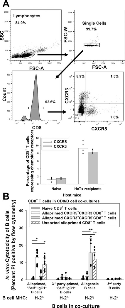
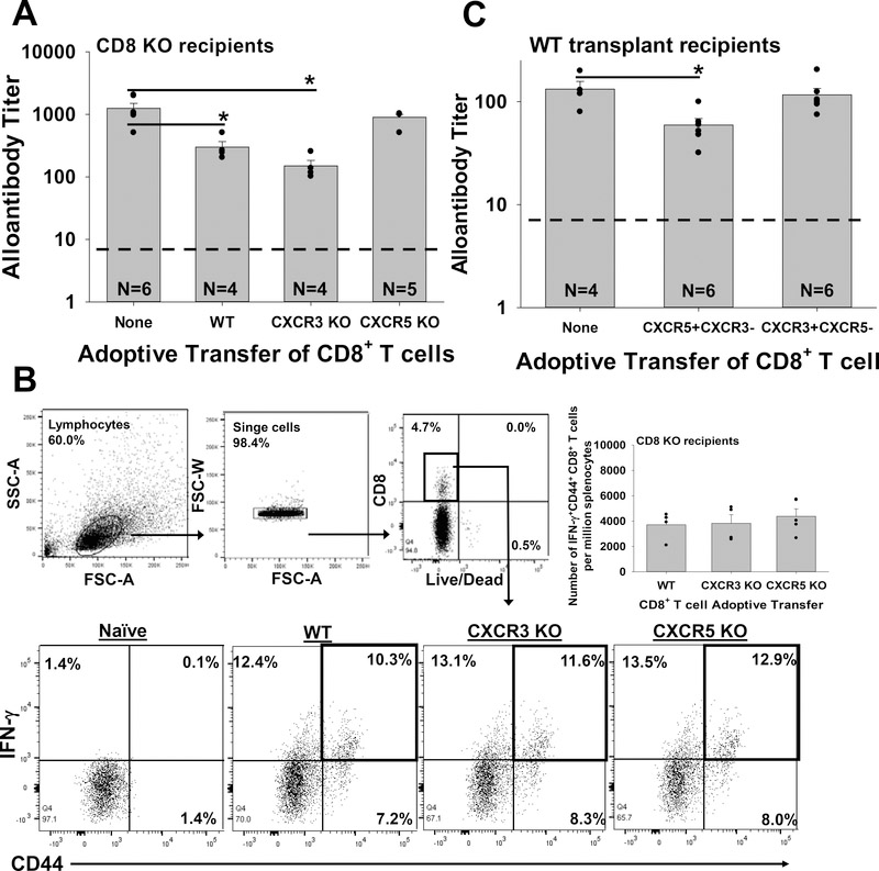
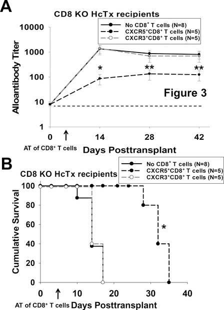
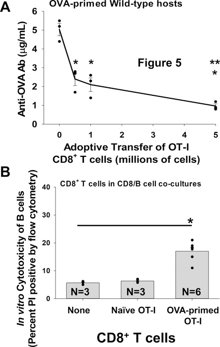
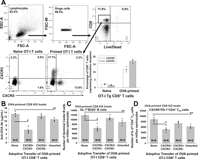
Methods: C57BL/6 mice were transplanted with FVB/N hepatocytes. Alloprimed CD8 T cells were retrieved on day 7 from hepatocyte transplant recipients. Unsorted or flow-sorted (CXCR5CXCR3 and CXCR3CXCR5) alloprimed CD8 T-cell subsets were analyzed for in vitro cytotoxicity and capacity to inhibit in vivo alloantibody production following adoptive transfer into C57BL/6 or high alloantibody-producing CD8 knock out (KO) hepatocyte transplant recipients. Alloantibody titer was assessed in CD8 KO mice reconstituted with naive CD8 T cells retrieved from C57BL/6, CXCR5 KO, or CXCR3 KO mice. Antibody suppression by ovalbumin (OVA)-primed monoclonal OVA-specific t-cell receptor transgenic CD8+ T cells (OT-I) CXCR5 or CXCR3 CD8 T-cell subsets was also investigated.

Results: Alloprimed CXCR5CXCR3CD8 T cells mediated in vitro cytotoxicity of alloprimed "self" B cells, while CXCR3CXCR5CD8 T cells did not. Only flow-sorted alloprimed CXCR5CXCR3CD8 T cells (not flow-sorted alloprimed CXCR3CXCR5CD8 T cells) suppressed alloantibody production and enhanced graft survival when transferred into transplant recipients. Unlike CD8 T cells from wild-type or CXCR3 KO mice, CD8 T cells from CXCR5 KO mice do not develop alloantibody-suppressor function. Similarly, only flow-sorted CXCR5CXCR3 (and not CXCR3CXCR5) OVA-primed OT-I CD8 T cells mediated in vivo suppression of anti-OVA antibody production.

Conclusions: These data support the conclusion that expression of CXCR5 by antigen-primed CD8 T cells is critical for the function of antibody-suppressor CD8 T cells.


Conflict of interest statement

Disclosures- The authors declare no conflicts of interest.

Figures

Figure 1.
Figure 1.. CXCR5+CD8+ T cells mediate in vitro cytotoxicity of alloprimed self IgG+ B cells.
C57BL/6 (wild-type, WT; H-2b) mice were transplanted with FVB/N (H-2q) hepatocytes. On day 7 post transplant, splenic CD8+ T cells were retrieved and purified. A) Flow cytometric analysis of purified CD8+ T cells shows that CXCR5+CXCR3 (9.3±1.0%) and CXCR3+CXCR5 (8.4±0.3%) CD8+ T cell subsets are detected (CD8+ T cells pooled from 10–15 mice) for cell sorting; data for n= 3 sorts are shown in the bar graph. B) In an in vitro cytotoxicity assay, flow-sorted alloprimed CXCR5+CXCR3 or CXCR3+CXCR5 CD8+ T cell populations and B cell targets were co-cultured at a 10:1 ratio for 4 hours and analyzed for cytotoxicity (propidium iodide (PI) uptake). Naïve CD8+ T cells and unsorted alloprimed CD8+ T cells were utilized as negative and positive controls (effector cells), respectively. Significant cytotoxicity of alloprimed, self IgG1+ B cells was observed in co-cultures with CXCR5+CXCR3CD8+ T cells (12.7±1.8%, n=10) or with unsorted alloprimed CD8+ T cells (9.1±3.8, p<0.0001 for both signified by “*”; n=10) in comparison to co-cultures with naïve CD8+ T cells (0.9±0.4%, n=9) but no significant cytotoxicity was detected in co-cultures with CXCR3+CXCR5CD8+ T cells (1.9±1.9%, p=ns, n=10). Significant cytotoxicity of allogeneic B cells was observed in co-cultures with CXCR3+CXCR5CD8+ T cells (15.6±8.8%; n=6) or with unsorted alloprimed CD8+ T cells (12.0±5.5%; n=6, p<0.002 for both signified by “**”), but not with CXCR5+CXCR3CD8+ T cells (1.8±1.8%; n=6, p=ns) in comparison to control co-cultures with naïve CD8+ T cells (0.8±0.7%; n=5). No significant cytotoxicity was detected against third-party (3rd) party primed “self” IgG1+ B cells (H-2b targets) or 3rd party B cells (H-2k targets) in any co-cultures. Error bars indicate standard error from triplicate experiments.
Figure 2.
Figure 2.. CXCR5 is critical to CD8+ T cell-mediated suppression of antibody production.
A) CD8 KO mice were transplanted with FVB/N hepatocytes. On day 0, recipients were adoptively transferred (AT) with 10×106 naïve CD8+ T cells (WT, CXCR5 KO or CXCR3 KO). Recipient serum was analyzed for alloantibody titer on day 14 post transplant. Significantly reduced alloantibody titer was observed in transplant mice that received AT of WT (300±70; n=4) or CXCR3 KO (150±30; n=4) CD8+ T cells (p<0.004 for both signified by “*”) compared to recipients that received AT with CXCR5 KO CD8+ T cells (900±100; n=5) or no AT (1,250±250; n=6). B) All groups of recipients that underwent transplant and adoptive transfer of WT, CXCR3 KO or CXCR5 KO CD8+ T cells developed alloprimed CD8+ T cells that were activated. Flow cytometric analysis gating on total leukocytes and CD8+ T revealed that by day 7 post transplant, approximately 10–13% of CD8+ T cells from all three groups (WT, CXCR3 KO, CXCR5 KO) of AT mice were IFN-γ+CD44+CD8+ T cells; similarly the quantity of IFN-γ+CD44+CD8+ T cells per million splenocytes was similar in all three groups. C) WT mice were transplanted with FVB/N hepatocytes and analyzed for production of alloantibody on day 14 post transplant. Transplant recipients that received AT (on day 5 post transplant) of flow-sorted (day 7) alloprimed CXCR5+CXCR3 WT CD8+ T cells exhibited significant reduction of alloantibody titer (60±10; n=5) compared to recipients which received AT of flow-sorted (day 7) alloprimed CXCR3+CXCR5CD8+ T cells (120±20; n=6) or no AT (130±20; n=4, p<0.02 for both signified by “*”). The dashed line on Figures 2A and 2C represents negative control sera from naïve mice. Error bars indicate standard error from duplicate experiments.
Figure 3.
Figure 3.. CXCR5+CD8+ T cell-mediated suppression of alloantibody enhances hepatocyte allograft survival.
CD8 KO mice were transplanted with FVB/N hepatocytes. On day 5, cohorts of transplant recipients received adoptive transfer of flow-sorted (day 7) alloprimed CXCR5+CXCR3CD8+ T cells, CXCR3+CXCR5CD8+ T cells, or no CD8+ T cells. Mice were observed for alloantibody production and hepatocyte survival. A) Adoptive transfer of CXCR5+CXCR3CD8+ T cells into CD8 KO recipients significantly inhibited alloantibody titer on day 14 (90±40; n=5, p<0.0001 signified by “*”) compared to no AT (1,300±500; n=8) while adoptive transfer of CXCR3+CXCR5CD8+ T cells did not (1,400±600; n=5, p=ns). Alloantibody titer remained suppressed after day 14 in CD8 KO mice that received AT of CXCR5+CXCR3CD8+ T cells (p<0.005 for days 28 and 42 post transplant, “**”). The dashed line represents negative control sera from naïve mice. Error bars indicate standard error. B) CD8 KO hepatocyte recipients that received adoptive transfer of CXCR5+CXCR3CD8+ T cells had prolonged allograft survival (MST=day 32; p=0.002, signified by “*”) compared to those that received AT of CXCR3+CXCR5CD8+ T cells (MST= day 14) or those with no AT (MST= day 14).
Figure 4.
Figure 4.. Suppression of alloantibody production by alloprimed CXCR5+CD8+ T cells is accompanied by a reduction in the quantity of Germinal Center B cells and CD4+ TFH cells.
WT mice were transplanted with FVB/N hepatocytes (day 0) and on day 5 received adoptive transfer (AT) of sorted alloprimed CXCR5+CXCR3CD8+ or CXCR3+CXCR5CD8+ T cells. On day 14 post transplant, recipient splenocytes were analyzed for the number of germinal center (GC) B cells and CD4+ TFH by flow cytometry. A) GC B cells (GL-7+B220+) were analyzed by gating on lymphocytes. B) Recipients which received AT of CXCR5+CXCR3CD8+ T cells exhibited significantly reduced number of GC B cells (22,000±2,000 per million splenocytes; n=5) compared to WT recipients without AT (38,000±2,000 per million splenocytes; n=5, p=0.001 signified by “*”). In contrast, the number of GC B cells was not significantly altered in recipients which received AT of CXCR3+CXCR5CD8+ T cells (41,000±3,000 per million splenocytes; n=6, p=ns) compared to control recipients without CD8+ T cell transfer. C) CD4+ TFH cells were analyzed by gating on lymphocytes, CD4+ T cells, and PD-1+CXCR5+ cells. D) Recipients which received AT of CXCR5+CXCR3CD8+ T cells exhibited significantly reduced number of IL-4+IL-21+ CD4+ TFH cells (4,400±100 per million splenocytes; n=5, p=0.003 signified by “*”) compared to WT recipients without AT (9,300±800 per million splenocytes; n=5). AT of CXCR5+CXCR3CD8+ T cells was associated with a reduced number of IL-4+ CD4+ TFH cells (2,400±100 per million splenocytes; n=5, p=0.04) compared to control recipients without CD8+ T cell transfer (4,600±500 per million splenocytes). In contrast, the number of IL-4+ (4,600±300 per million splenocytes; n=6) and IL-4+IL-21+ CD4+ TFH cells (8,100±500 per million splenocytes; n=6) was not significantly altered after AT of CXCR3+CXCR5CD8+ T cells compared to the control group with no CD8+ T cell transfer (p=ns for both IL-4+ and IL-4+IL-21+ CD4+ TFH cells). Error bars indicate standard error from duplicate experiments.
Figure 5.
Figure 5.. OVA-primed OT-I CD8+ T cells suppress anti-OVA antibody production in a dose-dependent manner and mediate in vitro cytotoxicity of OVA-primed B cells.
A) WT mice were primed with mOVA lysate (i.p. injection) on day 0. Cohorts of WT mice received adoptive transfer (AT) on day 0 of increasing quantities of OVA-primed OT-I TCR transgenic CD8+ T cells (0, 0.5, 1, 5×10⁶ cells i.v.). Mouse serum was assayed for anti-OVA antibodies (Ab) by ELISA on day 14 following antigen stimulation. WT mice primed with mOVA lysate exhibit maximal levels of serum anti-OVA antibodies on day 14 following antigen stimulation (5.0±0.3 μg/mL; n=3). AT of 0.5, 1, or 5×10⁶ OVA-primed OT-I CD8+ T cells on day 0 inhibited anti-OVA antibody production in these mice (2.4±0.4, 2.1±0.4, and 1.0±0.1 μg/mL respectively; n=3 and p<0.0004 for all groups signified by “*”). The AT of 5×10⁶ OVA-primed OT-I CD8+ T cells inhibited anti-OVA antibody production significantly more than 0.5 or 1×10⁶ OVA-primed OT-I CD8+ T cells (p<0.04 for both signified by “**”). B) OVA-primed OT-I CD8+ T cells or naïve OT-I CD8+ T cells were co-cultured with OVA-primed IgG1+ B cell targets. CD8+ T cells and B cells were co-cultured at a 10:1 ratio for 4 hours and analyzed for cytotoxicity. Significant cytotoxicity of OVA-primed IgG1+ B cells was observed in co-cultures with OVA-primed OT-I CD8+ T cells (17±1.4%; n=6, p<0.001 signified by “*”) compared to negative control cultures with no CD8+ T cells (5.7±0.3%) or with naïve OT-I CD8+ T cells (6.3±0.3%). Error bars indicate standard error.
Figure 6.
Figure 6.. OVA-primed OT-I CXCR5+CD8+ T cells mediate suppression of anti-OVA antibody production accompanied by reduced quantity of Germinal Center B cells and IL-21+CD4+ TFH cells.
A) Naïve OT-I CD8+ T cells were adoptively transferred (AT) into OVA-primed CD8 KO recipients. 7 days later, OT-I CD8+ T cells were isolated and analyzed by flow cytometry for expression of CXCR5 and CXCR3. A representative flow plot shows OVA-primed OT-I CD8+ T cells include CXCR5+CXCR3 (11.7±1.0%) and CXCR3+CXCR5 (17.2±0.7%) OT-I CD8+ T cell subsets. B) Unsorted or flow-sorted CXCR5+CXCR3 or CXCR3+CXCR5 OVA-primed OT-I CD8+ T cells (1×10⁶) were adoptively transferred into OVA-primed CD8 KO mice (day 5 post mOVA lysate stimulation). The amount of anti-OVA Ab produced in mice that received AT of CXCR5+CXCR3 OT-I CD8+ T cells (5.8±0.6 μg/mL; n=6, p<0.0001 signified by “*”) or unsorted OT-I CD8+ T cells (10.7±0.4 μg/mL, n=4, p=0.008 signified by “**”) was significantly less than the amount produced in CD8 KO recipients without AT of CD8+ T cells (13.4±0.3 μg/mL, n=6). AT of CXCR3+CXCR5 OT-I CD8+ T cells did not suppress anti-OVA antibody production (12.2±0.4 μg/mL; n=5, p=ns). C) The quantity of recipient spleen germinal center (GC) B cells (GL-7+B220+) and IL-21+CD4+ TFH cells (IL-21+CXCR5+PD-1+CD4+) was analyzed in OVA-primed CD8 KO mice (day 14 following mOVA lysate stimulation). The quantity of GC B cells in mice that received AT of CXCR5+CXCR3 OT-I CD8+ T cells (6,200±500 per million splenocytes; n=6, p<0.0001 signified by “*”) or with unsorted OT-I CD8+ T cells (8,900±700; n=4, p=0.008 signified by “**”) was significantly less than the quantity in CD8 KO recipients without AT (11,800±700 per million splenocytes; n=6). In contrast, AT of CXCR3+CXCR5 OT-I CD8+ T cells was not associated with reduction in the quantity of GC B cells (10,600±400 per million splenocytes; n=6, p=ns). D) The quantity of IL-21+ CD4+ TFH cells in mice that received AT of CXCR5+CXCR3 OT-I CD8+ T cells (5,000±400 per million splenocytes; n=6, p<0.0001 signified by “*”) or unsorted OT-I CD8+ T cells (6,100±300, n=4, p=0.001 signified by “**”) was significantly reduced compared to the quantity in CD8 KO recipients without AT (8,800±400 per million splenocytes; n=6). In contrast, AT with CXCR3+CXCR5 OT-I CD8+ T cells was not associated with a reduction in the quantity of IL-21+ CD4+ TFH cells (7,200±400 per million splenocytes; n=6, p=ns). Error bars indicate standard error from duplicate experiments.

Comment in

References

    1. Valenzuela NM, Reed EF. Antibody-mediated rejection across solid organ transplants: manifestations, mechanisms, and therapies. J Clin Invest. 2017;127(7):2492–2504. - PMC - PubMed
    1. Jorns C, Nowak G, Nemeth A, et al. De Novo Donor-Specific HLA Antibody Formation in Two Patients With Crigler-Najjar Syndrome Type I Following Human Hepatocyte Transplantation With Partial Hepatectomy Preconditioning. Am J Transplant. 2016;16(3):1021–1030. - PMC - PubMed
    1. Brooks AM, Carter V, Liew A, et al. De Novo Donor-Specific HLA Antibodies Are Associated With Rapid Loss of Graft Function Following Islet Transplantation in Type 1 Diabetes. Am J Transplant. 2015;15(12):3239–3246. - PubMed
    1. Heidt S, Roelen DL, Eijsink C, et al. Calcineurin inhibitors affect B cell antibody responses indirectly by interfering with T cell help. Clin Exp Immunol. 2010;159(2):199–207. - PMC - PubMed
    1. Blanchard D, Gaillard C, Hermann P, et al. Role of CD40 antigen and interleukin-2 in T cell-dependent human B lymphocyte growth. Eur J Immunol. 1994;24(2):330–335. - PubMed

Publication types

MeSH terms