. 2019 Feb 28;129(5):1984-1999.
doi: 10.1172/JCI122106. Print 2019 May 1.

Cleavage factor 25 deregulation contributes to pulmonary fibrosis through alternative polyadenylation

Affiliations

Cleavage factor 25 deregulation contributes to pulmonary fibrosis through alternative polyadenylation

Tingting Weng et al. J Clin Invest. .

Abstract

Idiopathic pulmonary fibrosis (IPF) is a chronic and deadly disease with a poor prognosis and few treatment options. Pathological remodeling of the extracellular matrix (ECM) by myofibroblasts is a key factor that drives disease pathogenesis, although the underlying mechanisms remain unknown. Alternative polyadenylation (APA) has recently been shown to play a major role in cellular responses to stress by driving the expression of fibrotic factors and ECMs through altering microRNA sensitivity, but a connection to IPF has not been established. Here, we demonstrate that CFIm25, a global regulator of APA, is down-regulated in the lungs of patients with IPF and mice with pulmonary fibrosis, with its expression selectively reduced in alpha-smooth muscle actin (α-SMA) positive fibroblasts. Following the knockdown of CFIm25 in normal human lung fibroblasts, we identified 808 genes with shortened 3'UTRs, including those involved in the transforming growth factor-β signaling pathway, the Wnt signaling pathway, and cancer pathways. The expression of key pro-fibrotic factors can be suppressed by CFIm25 overexpression in IPF fibroblasts. Finally, we demonstrate that deletion of CFIm25 in fibroblasts or myofibroblast precursors using either the Col1a1 or the Foxd1 promoter enhances pulmonary fibrosis after bleomycin exposure in mice. Taken together, our results identified CFIm25 down-regulation as a novel mechanism to elevate pro-fibrotic gene expression in pulmonary fibrosis.

Keywords: Fibrosis; Pulmonology.


Conflict of interest statement

Conflict of interest: The authors have declared that no conflict of interest exists.

Figures

Figure 1
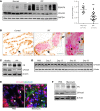
Figure 1. CFIm25 is downregulated in fibrotic lungs.
(A) A representative Western blot showing protein expression of CFIm25, FN, and GAPDH from 6 healthy and 7 IPF lung specimens. CFIm25 densitometric analysis shows significantly downregulated CFIm25 levels in IPF lungs. **P < 0.01, by unpaired t test versus healthy control. (B) Immunohistochemistry for CFIm25 (brown) and α-SMA (pink) showing cellular localization in control and IPF lung specimens. Arrow indicates CFIm25-positive cells. Arrowhead indicates α-SMA–positive but CFIm25-negative cells. Scale bars: 100 μm. Original magnification ×100. (C) Western blot shows CFIm25, FN, and β-actin protein expression levels in primary fibroblast lines derived from healthy or IPF lungs. (DF) Mice were i.p. injected with PBS or bleomycin biweekly for 4 weeks. (D) Western blotting was performed to analyze the protein expression of CFIm25 in whole-lung lysates on day 33 after PBS, or 7, 17, 28, and 33 days after the first bleomycin exposure. The different lanes represent samples collected from distinct mice. β-Actin was used as a protein loading control. (E) Immunofluorescence was carried out to determine CFIm25 (pink) and α-SMA (green) colocalization in lungs from mice exposed to PBS or bleomycin for 33 days. Arrows indicates CFIm25-positive cells; arrowheads indicatesα-SMA–positive but CFIm25-negative cells. Scale bars: 100 μm. (F) Primary fibroblasts were isolated from day-33 PBS or bleomycin-injected mouse lungs. Western blotting was performed to determine CFIm25, FN, and β-actin protein levels.
Figure 2
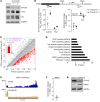
Figure 2. RNA-Seq identifies CFIm25 target genes involved in important fibrotic and cancer pathways.
(A) Western blot analysis of CFIm25, COL1, and FN expression in healthy human fibroblasts (CCD8-Lu) transfected with control siRNA (si-Con) or siRNAs targeting CFIm25 (si-CFIm25). GAPDH was used as a loading control. (B) Diagram shows pPAS and dPAS, and 2 sets of primers designed to target the translated region (P1) and distal region (P2) of the 3′-UTR. qRT-PCR was performed to demonstrate COL1A1 and FN1 transcript expression (lower left panel) and dPAS usage (lower right panel) after knockdown of CFIm25 using 2 different siRNAs (no. 1 and no. 2). Results in the lower left panel are shown as log2 (fold changes vs. control siRNA–transfected samples) ± SEM (n = 3 biological replicates), and results in the lower right panel are shown as the log2 ratio of (percentage of long transcript in si-CFIm25/percentage of long transcript in si-Con). *P < 0.05, by 1-sample t test versus 0. (C) Scatterplot of percentage PDUIs in control and CFIm25-knockdown cells, in which mRNAs were significantly shortened (n = 808) or lengthened (n = 29) after CFIm25 knockdown in CCD8-Lu cells. (D) Functional annotation assay of CFIm25 targets. (E) RNA-Seq read density for 3′-UTR, terminal exon, and upstream exon(s) of VMA21 in control or CFIm25 siRNA–transfected CCD8-Lu cells. Numbers on the x axis indicate the RNA-Seq read coverage. (F) qRT-PCR was performed to demonstrate dPAS usage of VMA21. n = 3. *P < 0.01, by 1-sample t test versus 0. (G) Western blotting was used to verify VMA21 expression after CFIm25 knockdown in CCD8-Lu cells. KD, knockdown.
Figure 3
Figure 3. TGF-β and Wnt5A pathways are activated in CFIm25-knockdown fibroblasts.
(A) The dPAS usage of CFIm25 targets involved in TGF-β (TGFBR1) and Wnt (WNT5A and FZD2) pathways was verified by qRT-PCR. n = 3 biological replicates. *P < 0.05 one sample t test versus 0. (B) RNA-Seq read density for a representative target (FZD2) is shown in control and CFIm25-knockdown CCD8-Lu cells. n = 3 biological replicates. P < 0.05, by 1-sample t test versus 0. Western blotting was performed to determine protein levels of (C) CFIm25 and TGF-βR1 and (D) Wnt5A and FZD2 in CFIm25-knockdown CCD8-Lu cells. (E) The dPAS usage of CFIm25 targets was determined using qRT-PCR in primary healthy (CCD8-Lu) or IPF fibroblasts (LL97A). n = 3 biological replicates. *P < 0.05, by 1-sample t test versus 0. (F and G) Western blotting was performed to determine protein levels of CFIm25, TGF-βR1, Wnt5A, and FZD2 in primary healthy (CCD8-Lu) and IPF fibroblasts (LL97A) (F) and IPF lungs with different levels of CFIm25 (G).
Figure 4
Figure 4. CFIm25 overexpression results in 3′-UTR lengthening and decreased protein expression of target genes.
Primary fibroblasts isolated from healthy and IPF lungs were electroporated with empty or CFIm25-overexpressing pCDNA3.1 plasmids. Two or three days after transfection, cells were collected for (A) Western blotting to determine the protein levels of CFIm25 and its target genes and (B) qRT-PCR to determine the dPAS usage of the CFIm25 targets COL1A1, TGFBR1, WNT5A, and FZD2. n = 3 biological replicates. *P < 0.05, by 1-sample t test versus 0.
Figure 5
Figure 5. Expression of CFIm25 and its targets in lung fibroblasts isolated from Col1a1-CreER-CFIm25fl/fl mice.
Four- to six-week-old Col1a1-CreER-CFIm25fl/fl mice and age- and sex-matched littermate controls were administrated 75 mg/kg (i.p.) tamoxifen daily for 5 days to induce Cre expression. n = 4 biological replications. One week later, fibroblasts were isolated from the lungs of these mice. (A) Western blotting was used to confirm the expression of CFIm25, Cre, and CFIm25 targets in fibroblasts, and (B) qRT-PCR was performed to determine the dPAS usage of CFIm25 targets (Col1a1, Tgfbr1, Fzd2, and Wnt5a). n = 5 biological replicates. *P < 0.05, by 1-sample t test versus 0.
Figure 6
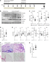
Figure 6. Bleomycin-induced pulmonary fibrosis is exaggerated in Col1a1-CreER-CFIm25fl/fl mice.
Four- to six-week-old Col1a1-CreER-CFIm25fl/fl mice and age- and sex-matched littermate controls were administrated 75 mg/kg tamoxifen (i.p.) daily for 5 days to induce Cre expression. After 1 week, the mice were injected with PBS or bleomycin biweekly for 4 weeks. Lungs were collected for analysis 28 days after the first bleomycin injection. (A) Western blot shows CFIm25, COL1, and FN expression in the lungs of control and Col1a1-CreER-CFIm25fl/fl mice. Pulmonary fibrosis was analyzed using a Sircol assay (B), qRT-PCR (C), Masson’s trichrome and α-SMA staining (D), and an Ashcroft assay (E). *P < 0.05, by 1-way ANOVA followed by Bonferroni’s multiple comparisons test versus Col1a1-CreER mice treated with bleomycin. n > 4 biological replicates.
Figure 7
Figure 7. Bleomycin-induced fibrosis is elevated in Col1a1-CreER-CFIm25fl/fl mice with delayed Cre activation.
(A) Diagram shows the experimental design. Four- to six-week-old Col1a1-CreER-CFIm25fl/fl mice and age- and sex-matched littermate controls were i.p. injected with PBS or bleomycin biweekly for 4 weeks. Starting on day 15 after the first bleomycin injection, the mice were i.p. injected with tamoxifen daily for 5 days to induce Cre recombination. Samples were collected for analysis 33 days after the first bleomycin injection. (B) Western blot shows CFIm25, Cre, COL1, FN, TGF-βR1, and Wnt5A expression in the lungs of control and Col1a1-CreER-CFIm25fl/+ mice. Levels of pulmonary fibrosis were analyzed using qRT-PCR (C) and a Sircol assay (D). (E) Lung function assay shows the parameters of tissue damping (G), tissue elastase (H), resistance (R), compliance (C), inspiratory capacity (IC), and Newtonian resistance (Rn). (F) Masson’s trichrome and α-SMA staining shows collagen deposition and myofibroblast differentiation. Scale bar: 200 μm. (G) The pulmonary fibrosis seen in Masson’s trichrome–stained slides was quantified by blinded Ashcroft assay. *P < 0.05, by unpaired t test with equal variance. n = 8 biological replicates.
Figure 8
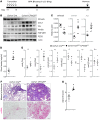
Figure 8. Col1a1-CreER-CFIm25fl/fl mice have more severe pulmonary fibrosis upon o.p.a. bleomycin injection.
(A) Diagram shows the mouse treatment timeline. Four- to six-week-old Col1a1-CreER-CFIm25fl/fl mice and age- and sex-matched littermate controls were administrated 75 mg/kg tamoxifen (i.p.) daily for 5 days to induce Cre activation. After 1 week, mice were injected with PBS or bleomycin through o.p.a. instillation. Lungs were collected for analysis 21 days after the first bleomycin injection. (B) Western blot shows CFIm25, COL1, FN, TGF-βR1, Wnt5A, and FZD2 expression in the lungs of control and Col1a1-CreER-CFIm25fl/+ mice. qRT-PCR (C) and a Sircol assay (D) were carried out to analyze the levels of pulmonary fibrosis. (E) Tissue damping, tissue elastase, resistance, compliance, inspiratory capacity, and Newtonian resistance were analyzed using the flexiVent system. (F) Masson’s trichrome and α-SMA staining indicated collagen deposition and myofibroblast differentiation. Scale bars: 200 μm. (G) Blinded Ashcroft assays were performed to quantify the pulmonary fibrosis observed on Masson’s trichrome–stained slides. *P < 0.05, by unpaired t test with equal variance. n > 4 biological replicates.

References

    1. Steele MP, Schwartz DA. Molecular mechanisms in progressive idiopathic pulmonary fibrosis. Annu Rev Med. 2013;64:265–276. doi: 10.1146/annurev-med-042711-142004. - DOI - PubMed
    1. Wolters PJ, Collard HR, Jones KD. Pathogenesis of idiopathic pulmonary fibrosis. Annu Rev Pathol. 2014;9:157–179. doi: 10.1146/annurev-pathol-012513-104706. - DOI - PMC - PubMed
    1. Chanda D, Otoupalova E, Smith SR, Volckaert T, De Langhe SP, Thannickal VJ. Developmental pathways in the pathogenesis of lung fibrosis. Mol Aspects Med. 2019;65:56–69. doi: 10.1016/j.mam.2018.08.004. - DOI - PMC - PubMed
    1. Zank DC, Bueno M, Mora AL, Rojas M. Idiopathic pulmonary fibrosis: aging, mitochondrial dysfunction, and cellular bioenergetics. Front Med (Lausanne) 2018;5:10. - PMC - PubMed
    1. Todd NW, Luzina IG, Atamas SP. Molecular and cellular mechanisms of pulmonary fibrosis. Fibrogenesis Tissue Repair. 2012;5(1):11. doi: 10.1186/1755-1536-5-11. - DOI - PMC - PubMed

Publication types

MeSH terms