. 2019 Jun;176(11):1728-1744.
doi: 10.1111/bph.14647. Epub 2019 Apr 15.

The specialised pro-resolving lipid mediator maresin 1 reduces inflammatory pain with a long-lasting analgesic effect

Affiliations

The specialised pro-resolving lipid mediator maresin 1 reduces inflammatory pain with a long-lasting analgesic effect

Victor Fattori et al. Br J Pharmacol. 2019 Jun.

Abstract

Background and purpose: Maresin 1 (MaR1) is a specialised pro-resolving lipid mediator with anti-inflammatory and analgesic activities. In this study, we addressed the modulation of peripheral and spinal cord cells by MaR1 in the context of inflammatory pain.

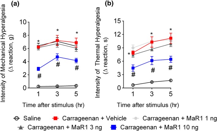
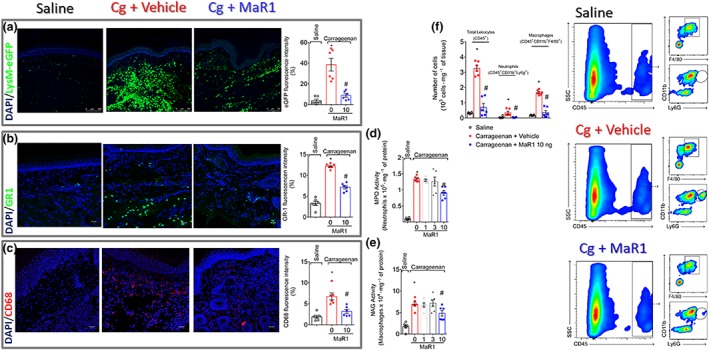
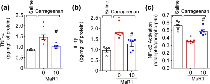
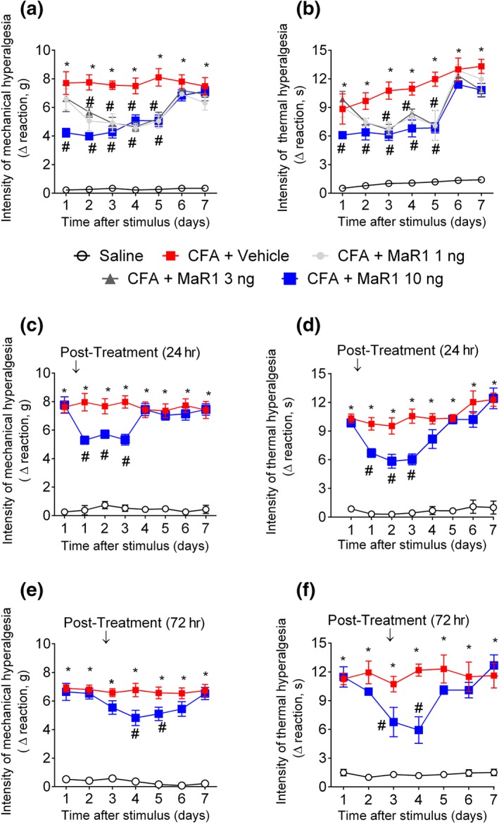
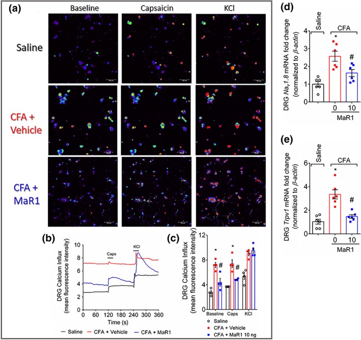
Experimental approach: Mice were treated with MaR1 before intraplantar injection of carrageenan or complete Freund's adjuvant (CFA). Mechanical hyperalgesia was assessed using the electronic von Frey and thermal hyperalgesia using a hot plate. Spinal cytokine production and NF-κB activation were determined by ELISA and astrocytes and microglia activation by RT-qPCR and immunofluorescence. CGRP release by dorsal root ganglia (DRG) neurons was determined by EIA. Neutrophil and macrophage recruitment were determined by immunofluorescence, flow cytometry, and colorimetric methods. Trpv1 and Nav1.8 expression and calcium imaging of DRG neurons were determined by RT-qPCR and Fluo-4AM respectively.

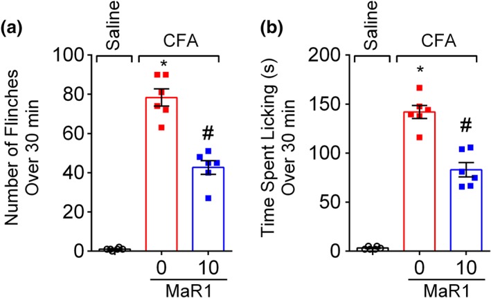
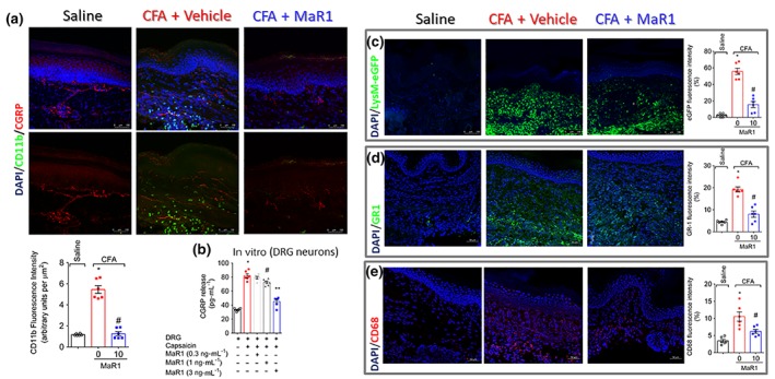
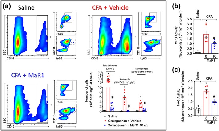
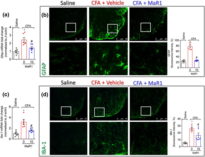
Key results: MaR1 reduced carrageenan- and CFA-induced mechanical and thermal hyperalgesia and neutrophil and macrophage recruitment proximal to CGRP+ fibres in the paw skin. Moreover, MaR1 reduced NF-κB activation, IL-1β and TNF-α production, and spinal cord glial cells activation. In the DRG, MaR1 reduced CFA-induced Nav1.8 and Trpv1 mRNA expression and calcium influx and capsaicin-induced release of CGRP by DRG neurons.

Conclusions and implications: MaR1 reduced DRG neurons activation and CGRP release explaining, at least in part, its analgesic and anti-inflammatory effects. The enduring analgesic and anti-inflammatory effects and also post-treatment activity of MaR1 suggest that specialised pro-resolving lipid mediators have potential as a new class of drugs for the treatment of inflammatory pain.


Conflict of interest statement

The authors declare no conflicts of interest.

Figures

Figure 1
Figure 1
MaR1 reduces carrageenan‐induced mechanical and thermal hyperalgesia. Mechanical hyperalgesia (a) and thermal hyperalgesia (b) were evaluated 1, 3, and 5 hr after intraplantar injection of carrageenan (100 μg per paw). Results from mechanical hyperalgesia are presented as Δ withdrawal threshold (in g) and for thermal hyperalgesia as Δ withdrawal threshold (in s), which was calculated by subtracting the mean measurements at 1–5 hr after carrageenan from the zero‐time (baseline values) mean measurements. MaR1 reduced carrageenan‐induced mechanical (a) and thermal (b) hyperalgesia. Results are representative of two independent experiments and are presented as mean ± SEM of measurements, n = 6 mice per group per experiment (*P < 0.05 vs. saline, # P < 0.05 vs. 0 mg·kg−1 group; two‐way repeated measures ANOVA followed by Tukey's post‐test)
Figure 2
Figure 2
MaR1 inhibits carrageenan‐induced neutrophil and macrophage recruitment to the hind paw skin. Hind paw skin of LysM‐eGFP (neutrophil and macrophage marker, a) or Swiss (b–f) mice was dissected for determination of neutrophil (GR‐1 staining [b], MPO activity [d], and flow cytometry [CD11b+Ly6G+ cells, f]) and macrophage (CD68 staining [c], NAG activity [e], and flow cytometry [CD11b+F4/80+ cells, f]) recruitment 5 hr after carrageenan stimulus. LysM‐eGFP mouse, immunofluorescence, enzymatic activity, and flow cytometry data show that MaR1 reduced carrageenan‐induced recruitment of neutrophils and macrophages in the paw skin. Results are expressed as mean ± SEM, n = 6 mice per group per experiment, two independent experiments (*P < 0.05 vs. saline, # P < 0.05 vs. 0 mg·kg−1 group; one‐way ANOVA followed by Tukey's post‐test)
Figure 3
Figure 3
MaR1 inhibits carrageenan‐induced spinal cord cytokine production and NF‐κB activation. Three hours after intraplantar injection of carrageenan (100 μg per paw), the spinal cord was dissected for determination of TNF‐α (a), IL‐1β (b), and NF‐κB activation (c) by ELISA. NF‐κB activation was observed as a reduction of total p65/phosphorylated p65 OD ratio. Results are representative of two independent experiments and are presented as mean ± SEM, n = 6 mice per group per experiment (*P < 0.05 vs. saline, # P < 0.05 vs. 0 mg·kg−1 group; one‐way ANOVA followed by Tukey's post‐test)
Figure 4
Figure 4
MaR1 reduces CFA‐induced mechanical and thermal hyperalgesia. Mechanical hyperalgesia (a, c, and e) and thermal hyperalgesia (b, d, and f) were evaluated 1–7 days after intraplantar injection of CFA (10 μl per paw). Results from mechanical hyperalgesia are presented as Δ withdrawal threshold (in g) and for thermal hyperalgesia as Δ withdrawal threshold (in s), which were calculated by subtracting the mean measurements at 1–7 days after carrageenan from the zero‐time (baseline values) mean measurements. Panels (a) and (b) show the analgesic effect of MaR1 as a 20 min pretreatment. Panels (c) and (d) show the analgesic effect of MaR1 as a post‐treatment 1 day after CFA. The measurements on the first day (c and d) were shown before and after treatment. Panels (e) and (f) show the analgesic effect of MaR1 as a post‐treatment 3 days after CFA. Results are representative of two independent experiments and are presented as mean ± SEM of measurements, n = 6 mice per group per experiment (*P < 0.05 vs. saline, # P < 0.05 vs. 0 mg·kg−1 group; two‐way repeated measures ANOVA followed by Tukey's post‐test)
Figure 5
Figure 5
MaR1 decreases CFA‐induced overt pain‐like behaviour. CFA induced repetitive paw flinches (a) and licking of the paw (b), which were determined over 30 min 1 day after intraplantar injection of CFA (10 μl per paw). Results are representative of two independent experiments and are presented as mean ± SEM, n = 6 mice per group per experiments (*P < 0.05 vs. saline, # P < 0.05 vs. 0 mg·kg−1 group; one‐way ANOVA followed by Tukey's post‐test)
Figure 6
Figure 6
MaR1 reduces the number of leukocytes proximal to CGRP+ fibres and the release of CGRP by DRG neurons. Hind paw skin was dissected for determination of total leukocytes (CD11b+ cells) close to CGRP+ fibres (a), which showed an increase of total CD11b fluorescence proximal to CGRP+ fibres in the CFA group and reduction after MaR1 treatment. For CGRP release assay (b), naïve DRG neurons received vehicle or different concentrations of MaR1 (0.3, 1, or 3 ng·ml−1) before stimulus with capsaicin. Supernatant was collected 1 hr after capsaicin to determine CGRP levels by EIA. Panels (c) to (e) analysed with further detail the cellular types recruited to the paw skin during CFA inflammation. LysM‐eGFP (C57BL/6 background mice) was used to determine neutrophils and macrophages (c). The staining neutrophils (GR‐1, d) and macrophages (CD68, e) in hind paw skin samples of Swiss mice also showed that MaR1 reduced CFA‐induced recruitment of these cells. Results are expressed as mean ± SEM, n = 6 mice per group per experiment, two independent experiments (*P < 0.05 vs. saline, # P < 0.05 vs. 0 mg·kg−1 group; one‐way ANOVA followed by Tukey's post‐test). Results are expressed as mean ± SEM, n = 6 wells per group per experiment, two independent experiments (*P < 0.05 vs. saline, # P < 0.05 vs. vehicle group; ** P < 0.05 vs. 1 ng·ml−1 group; one‐way ANOVA followed by Tukey's post‐test)
Figure 7
Figure 7
MaR1 inhibits CFA‐induced CD11b+Ly6G+ neutrophils and CD11b+F4/80+ macrophage recruitment to the hind paw skin. Hind paw skin was dissected for determination of neutrophil (flow cytometry [a] and MPO activity [b]) and macrophage recruitment (flow cytometry [a] and NAG activity [c]) 3 days after the stimulus. Results are expressed as mean ± SEM, n = 6 mice per group per experiment, two independent experiments (* P < 0.05 vs. saline, # P < 0.05 vs. 0 mg·kg−1 group; one‐way ANOVA followed by Tukey's post‐test)
Figure 8
Figure 8
MaR1 inhibits CFA‐induced spinal cord cytokine production and NF‐κB activation. Three days after intraplantar injection of CFA (10 μl per paw), the spinal cord was dissected for determination of TNF‐α (a), IL‐1β (b), and NF‐κB activation (c) by ELISA. NF‐κB activation was observed as a reduction of total p65/phosphorylated p65 OD ratio. Results are representative of two independent experiments and are presented as mean ± SEM, n = 6 mice per group per experiments (*P < 0.05 vs. saline, # P < 0.05 vs. 0 mg·kg−1 group; one‐way ANOVA followed by Tukey's post‐test)
Figure 9
Figure 9
MaR1 decreases CFA‐induced astrocyte and microglia activation. Three days after intraplantar injection of CFA (10 μl per paw), spinal cord was dissected for determination of astrocyte and microglia activation by RT‐qPCR (a and c) and by immunofluorescence (b and d). Glial fibrillary acidic protein (GFAP) was used as a marker of the activation of astrocytes (a and b), and IBA‐1 was used as a marker of microglia activation (c and d). Results are representative of two independent experiments and are presented as mean ± SEM, n = 6 mice per group per experiments (*P < 0.05 vs. saline, # P < 0.05 vs. 0 mg·kg−1 group, **P < 0.05 vs. 10 mg·kg−1; one‐way ANOVA followed by Tukey's post‐test)
Figure 10
Figure 10
MaR1 reduces CFA‐induced activation of DRG neurons. Three days after intraplantar injection of CFA (10 μl per paw), DRGs were dissected for calcium imaging using Fluo‐4AM (a–c) and mRNA expression by RT‐qPCR (d and e). Panel (a) displays representative fields of DRG neurons dissected from saline‐treated mice, mice stimulated with CFA and treated with vehicle, or stimulated with CFA and treated with MaR1. Panel (a): baseline fluorescence (first column), fluorescence after capsaicin (second column), and after KCl control (third column). Panel (b) displays the fluorescence intensity traces of calcium influx from the representative DRG fields (a) throughout the 6 min of recording. The representative traces show that the CFA + vehicle DRG neurons presented higher calcium levels in the baseline than saline control and CFA + MaR1 DRG neurons groups. Panel (c) shows the mean fluorescence intensity of calcium influx of the baseline (0‐s mark) and that following the stimulus, either capsaicin (120‐s mark, TRPV1 agonist) or KCl (240‐s mark, activates all neurons). Panels (d) and (e) show the DRG neurons RT‐qPCR data demonstrating that MaR1 reduced CFA‐induced Nav1.8 (d) and Trpv1 (e) mRNA expression. Results are expressed as mean ± SEM, n = 4 DRG plates (each plate is a neuronal culture pooled from six mice) per group per experiment, and RT‐qPCR used n = 6 DRG per group per experiment, two independent experiments (*P < 0.05 vs. saline, # P < 0.05 vs. 0 mg·kg−1 group; one‐way ANO XVA followed by Tukey's post‐test)

Similar articles

Cited by

References

    1. Alexander, S. P. , Christopoulos, A. , Davenport, A. P. , Kelly, E. , Marrion, N. V. , Peters, J. A. , … CGTP Collaborators . (2017). The Concise Guide to PHARMACOLOGY 2017/18: G protein‐coupled receptors. British Journal of Pharmacology, 174(Suppl 1), S17–S129. 10.1111/bph.13878 - DOI - PMC - PubMed
    1. Alexander, S. P. H. , Fabbro, D. , Kelly, E. , Marrion, N. V. , Peters, J. A. , Faccenda, E. , … CGTP Collaborators . (2017). The Concise Guide to PHARMACOLOGY 2017/18: Enzymes. British Journal of Pharmacology, 174(S1), S272–S359. 10.1111/bph.13877 - DOI - PMC - PubMed
    1. Alexander, S. P. , Striessnig, J. , Kelly, E. , Marrion, N. V. , Peters, J. A. , Faccenda, E. , … CGTP Collaborators . (2017). The Concise Guide to PHARMACOLOGY 2017/18: Voltage‐gated ion channels. British Journal of Pharmacology, 174(Suppl 1), S160–S194. 10.1111/bph.13884 - DOI - PMC - PubMed
    1. Alexandre, C. , Latremoliere, A. , Ferreira, A. , Miracca, G. , Yamamoto, M. , Scammell, T. E. , & Woolf, C. J. (2017). Decreased alertness due to sleep loss increases pain sensitivity in mice. Nature Medicine, 23(6), 768–774. 10.1038/nm.4329 - DOI - PMC - PubMed
    1. Bannenberg, G. L. , Chiang, N. , Ariel, A. , Arita, M. , Tjonahen, E. , Gotlinger, K. H. , … Serhan, C. N. (2005). Molecular circuits of resolution: Formation and actions of resolvins and protectins. Journal of Immunology, 174(7), 4345–4355. 10.4049/jimmunol.174.7.4345 - DOI - PubMed

Publication types

MeSH terms