Review
. 2019 Apr:375:14-24.
doi: 10.1016/j.heares.2019.02.007. Epub 2019 Feb 22.

Usher syndrome and non-syndromic deafness: Functions of different whirlin isoforms in the cochlea, vestibular organs, and retina

Affiliations
Review

Usher syndrome and non-syndromic deafness: Functions of different whirlin isoforms in the cochlea, vestibular organs, and retina

Pranav Dinesh Mathur et al. Hear Res. 2019 Apr.

Abstract

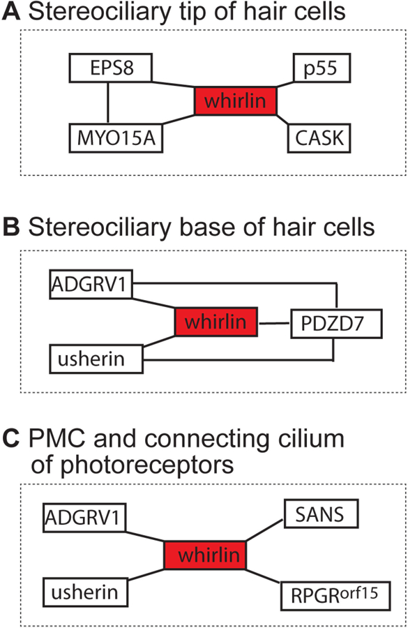
Usher syndrome (USH) is the leading cause of inherited combined vision and hearing loss. However, mutations in most USH causative genes lead to other diseases, such as hearing loss only or vision loss only. The molecular mechanisms underlying the variable disease manifestations associated with USH gene mutations are unclear. This review focuses on an USH type 2 (USH2) gene encoding whirlin (WHRN; previously known as DFNB31), mutations in which have been found to cause either USH2 subtype USH2D or autosomal recessive non-syndromic deafness type 31 (DFNB31). This review summarizes the current knowledge about different whirlin isoforms encoded by WHRN orthologs in animal models, the interactions of different whirlin isoforms with their partners, and the function of whirlin isoforms in different cellular and subcellular locations. The recent findings regarding the function of whirlin isoforms suggest that disruption of different isoforms may be one of the mechanisms underlying the variable disease manifestations caused by USH gene mutations. This review also presents recent findings about the vestibular defects in Whrn mutant mouse models, which suggests that previous assumptions about the normal vestibular function of USH2 patients need to be re-evaluated. Finally, this review describes recent progress in developing therapeutics for diseases caused by WHRN mutations.

Keywords: Ankle link; Hair cell; Hearing loss; Photoreceptor; Retinitis pigmentosa; Stereocilia; Usher syndrome.


Conflict of interest statement

Declarations of interest: none

Figures

Figure 1:
Figure 1:
Structure of an inner ear hair cell and a retinal photoreceptor. (A) A developing cochlear hair cell (e.g., mouse P2–P12) has ankle links (highlighted in blue) located at the base of stereocilia. Lateral links and horizontal top connectors are also present. Tip links connect the tip of short stereocilia to the shaft of longer stereocilia in the adjacent row and are required for mechanotransduction. (B) A rod photoreceptor has an outer segment with discs that contain rhodopsin. Rhodopsin detects light in the form of photons. The photoreceptor inner segment contains mainly organelles required for energy metabolism and protein synthesis. The periciliary membrane complex (highlighted in blue) is located at the apical region of inner segment, surrounding the connecting cilium (highlighted in orange). In order to illustrate the periciliary membrane complex and the connecting cilium, the calyceal processes, which are located outside the two subcellular structures, are not shown. The cell body contains the nucleus, while the synaptic region contains ribbon synapses.
Figure 2:
Figure 2:
Location and disease manifestation of human WHRN mutations. FL-whirlin mRNA (untranslated region-light blue; coding sequence region-green) is translated into a protein with two harmonin N-like (HNL) domains, three PDZ domains, a proline-rich (PR) region, and a PDZ-binding motif (PBM). Arabic numerals in the coding sequence region denote exon numbers. The asterisks and short line in red denote mutations in the N-terminal region that cause USH2D, except p.Q54X. The patient with p.Q54X mutation has not been tested for hearing. The asterisks in blue denote C-terminal mutations that cause DFNB31.
Figure 3:
Figure 3:
Whrn genomic DNA and mRNA variants in wild-type and Whrn mutant mice. (A) FL-, N-, and C-terminal whirlin mRNA variants found in the wild-type mice. For simplicity, only one representative N-whirlin mRNA variant is shown, although several N-whirlin mRNA variants have been reported (Belyantseva et al., 2005; Ebrahim et al., 2016; Mathur et al., 2015b; Wright et al., 2012). In addition, the C-whirlin variants starting from exon 1 are not shown. (B) A deletion from exon 6 to exon 10 (red box) truncates both FL-whirlin mRNA and C-whirlin mRNA variants in Whrnwi mice. A vertical yellow bar indicates a premature stop in truncated Whrnwi mRNA fragments. N-whirlin mRNA variants were not found in our study, although they were found in the Whrnwi inner ear by Ebrahim et al. (Ebrahim et al., 2016; Mathur et al., 2015b; Mathur et al., 2015c). (C) Only a C-terminal whirlin variant is present in Whrnneo mice, which has a Neor cassette (orange box) in the 5′ region of exon 1. (D) A targeting cassette (light blue box) in intron 3 of Whrntm1a genomic DNA (gDNA) disrupts the FL-whirlin variant. However, it is expected that the C-terminal whirlin mRNA variant remains unaffected. L, loxP site. (E) Exon 4 and a part of the targeting cassette (light blue box) in Whrntm1a gDNA are deleted in Whrntm1b mice, which disrupts the FL-whirlin variant. The C-terminal whirlin mRNA variant is expected to be unaffected. (F) Whrnwi+BAC mice are expected to express C-terminal whirlin mRNA variant from the transgenic BAC DNA, in addition to the variants expressed in Whrnwi mice. Brown and light grey in the Whrn gDNA and BAC DNA denote the exons and introns, respectively, with exon numbers in Arabic numerals. White color denotes 5′ and 3′-untranslated regions, and green color denotes the coding sequence region of whirlin mRNA variants.
Figure 4:
Figure 4:
Localization of whirlin isoforms in the wild-type and Whrn mutant inner ear and retina. (A) In the developing wild-type organ of Corti (e.g., during mouse P2–P12), FL-whirlin is localized to the stereociliary base of IHCs and OHCs. Additionally, IHC stereociliary tips contain FL- and C-whirlin, while OHC stereociliary tips contain only C-whirlin. (B) Localization of whirlin isoforms in wild-type VHCs is similar to that of developing IHCs as shown in A. (C) FL- and C-whirlin isoforms remain at the stereociliary tip of mature wild-type IHCs, but no whirlin isoforms are present in mature wild-type OHCs. (D) FL-whirlin and likely N-whirlin are localized to the PMC in wild-type photoreceptor cells. (E-G) C-whirlin is localized to the stereociliary tip of OHCs, IHCs, and VHCs in Whrnneo mice. This localization of C-whirlin in OHCs occurs only during development but not in adulthood. No whirlin isoforms are present at the stereociliary base in any Whrnneo hair cells at any time point. (H) No whirlin isoforms are detected in Whrnneo photoreceptors. (I-K) Whrnwi inner ear hair cells do not show any whirlin expression at any time point. (L) The truncated N-whirlin fragment in Whrnwi retinas localizes to the PMC. (M-N) 96% of IHCs and 33% of OHCs in Whrnwi+BAC mice show localization of C-whirlin to their stereociliary tips during development. (O) Similar to Whrnneo mouse, Whrntm1a and Whrntm1b mice show localization of only C-whirlin to their IHC and OHC stereociliary tips during development. In this figure, the short and thick stereocilia phenotypes in Whrnneo and Whrnwi hair cells are shown.
Figure 5:
Figure 5:
Interacting partners of whirlin at various subcellular locations in the inner ear and retina. (A) Whirlin interacts with EPS8, MYO15A, p55, and CASK proteins at the stereociliary tip of inner ear hair cells, where MYO15A and EPS8 also interact with each other. (B) Whirlin interacts with ADGRV1, usherin, and PDZD7 at the stereociliary base in inner ear hair cells, where the four proteins interact among one another and form the ALC. (C) Whirlin interacts with ADGRV1 and usherin at the PMC and with SANS and RPGRorf15 at the connecting cilium in photoreceptors.

References

    1. Audo I, Bujakowska K, Mohand-Said S, Tronche S, Lancelot ME, Antonio A, Germain A, Lonjou C, Carpentier W, Sahel JA, Bhattacharya S, Zeitz C 2011. A novel DFNB31 mutation associated with Usher type 2 syndrome showing variable degrees of auditory loss in a consanguineous Portuguese family. Mol. Vis 17, 1598–606. - PMC - PubMed
    1. Belyantseva IA, Boger ET, Naz S, Frolenkov GI, Sellers JR, Ahmed ZM, Griffith AJ, Friedman TB 2005. Myosin-XVa is required for tip localization of whirlin and differential elongation of hair-cell stereocilia. Nat Cell Biol 7, 148–56. - PubMed
    1. Besnard T, Vache C, Baux D, Larrieu L, Abadie C, Blanchet C, Odent S, Blanchet P, Calvas P, Hamel C, Dollfus H, Lina-Granade G, Lespinasse J, David A, Isidor B, Morin G, Malcolm S, Tuffery-Giraud S, Claustres M, Roux AF 2012. Non-USH2A mutations in USH2 patients. Hum. Mutat 33, 504–10. - PubMed
    1. Boughman JA, Vernon M, Shaver KA 1983. Usher syndrome: definition and estimate of prevalence from two high-risk populations. J. Chronic Dis 36, 595–603. - PubMed
    1. Chen Q, Zou J, Shen Z, Zhang W, Yang J 2014. Whirlin and PDZ domain containing 7 (PDZD7) proteins are both required to form the quaternary protein complex associated with Usher syndrome type 2. J. Biol. Chem 289, 36070–36088. - PMC - PubMed

Publication types

MeSH terms

Supplementary concepts