

Idiopathic intracranial hypertension presenting as bilateral spontaneous lateral intrasphenoidal and transethmoidal meningoceles: a case report and review of the literature
- PMID: 30832738
- PMCID: PMC6399895
- DOI: 10.1186/s13256-018-1959-6
Idiopathic intracranial hypertension presenting as bilateral spontaneous lateral intrasphenoidal and transethmoidal meningoceles: a case report and review of the literature
Abstract
Background: Basal meningoceles are rare herniations of the meninges that tend to present unilaterally with cerebrospinal fluid rhinorrhea. Growing evidence suggests that intracranial hypertension contributes considerably to the formation of spontaneous basal meningoceles.
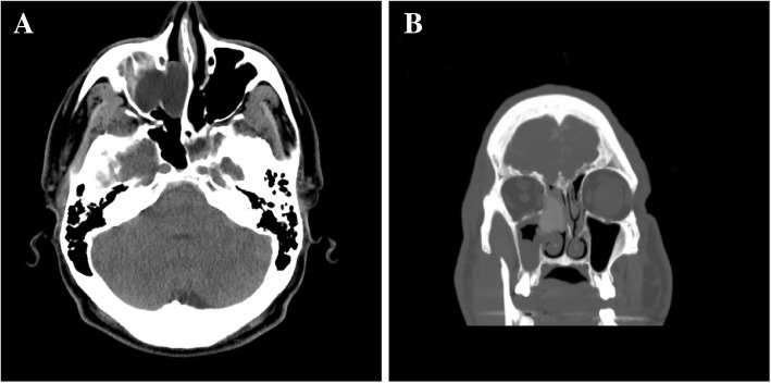
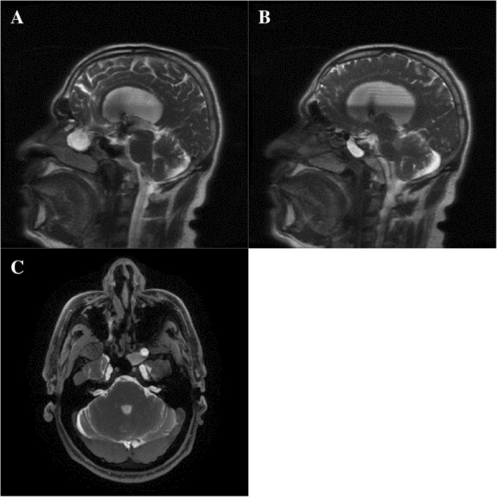
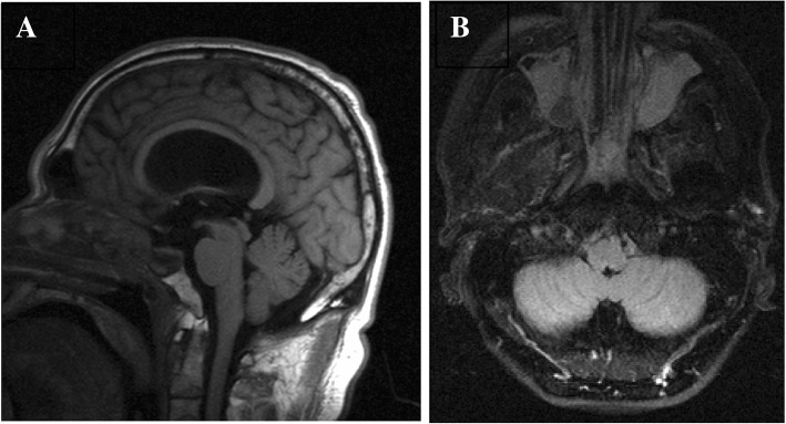
Case presentation: A 50-year-old man of Middle East ethnicity presented with a 16-week history of cerebrospinal fluid rhinorrhea, short-term memory loss, and slight decline in cognitive function. We present a case of bilateral spontaneous meningoceles with bone defects in the left lateral sphenoid sinus and right anterior cribriform plate, as well as with a remodeled sella. A neuronavigation-assisted expanded endoscopic endonasal surgery was performed to resect the meningoceles. Postoperative imaging demonstrated complete resolution of the bilateral meningoceles.
Conclusions: This case reports the first bilateral basal spontaneous meningoceles in the literature. Furthermore, based on this case's imaging results and the literature reviewed, elevated intracranial pressure may be a determining factor behind the development of spontaneous meningoceles.
Keywords: Bilateral; Case report; Endonasal; Expanded endoscopic; Lateral intrasphenoidal; Meningocele; Skull base; Spontaneous; Surgery; Transethmoidal.
Conflict of interest statement
Ethics approval and consent to participate
Not applicable.
Consent for publication
Written informed consent was obtained from the patient for publication of this case report and any accompanying images. A copy of the written consent is available for review by the Editor-in-Chief of this journal.
Competing interests
The authors declare that they have no competing interests.
Publisher’s Note
Springer Nature remains neutral with regard to jurisdictional claims in published maps and institutional affiliations.
Figures
References
Publication types
MeSH terms
LinkOut - more resources
Full Text Sources
