. 2019 May 8;27(5):922-932.
doi: 10.1016/j.ymthe.2019.02.011. Epub 2019 Feb 16.

Adipocytes: A Novel Target for IL-15/IL-15Rα Cancer Gene Therapy

Affiliations

Adipocytes: A Novel Target for IL-15/IL-15Rα Cancer Gene Therapy

Run Xiao et al. Mol Ther. .

Abstract

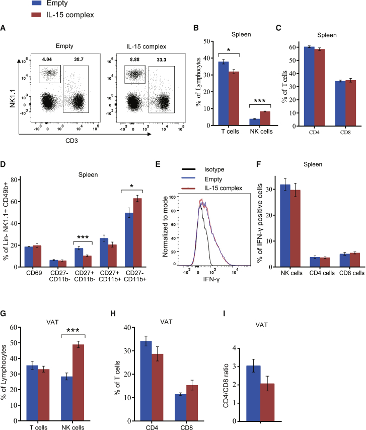
IL-15 is a proinflammatory cytokine that plays an essential role in the development and activation of natural killer (NK) cells. Adipose tissue acts as an endocrine organ that secretes cytokines and is an important reservoir for lymphocytes. We hypothesized that activation of the IL-15 signaling in adipose tissue will activate and expand the NK cell population and control tumor growth. We recently developed an adipocyte-targeting recombinant adeno-associated viral (rAAV) vector with minimal off-target transgene expression in the liver. Here, we used this rAAV system to deliver an IL-15/IL-15Rα complex to the abdominal fat by intraperitoneal (i.p.) injection. Adipose IL-15/IL-15Rα complex gene transfer led to the expansion of NK cells in the adipose tissue and spleen in normal mice without notable side effects. The i.p. injection of rAAV-IL-15/IL-15Rα complex significantly suppressed the growth of Lewis lung carcinoma implanted subcutaneously and exerted a significant survival advantage in a B16-F10 melanoma metastasis model. The antitumor effects were associated with the expansion of the NK cells in the blood, spleen, abdominal fat, and tumor, as well as the enhancement of NK cell maturity. Our proof-of-concept preclinical studies demonstrate the safety and efficacy of the adipocyte-specific IL-15/IL-15Rα complex vector as a novel cancer immune gene therapy.

Keywords: IL-15/IL-15Rα; NK cell; adipocyte; cancer; gene therapy; immune therapy; rAAV; visceral fat.


Figures

None
Graphical abstract
Figure 1
Figure 1
Adipocyte Gene Transfer of the IL-15/IL-15Rα Complex Shows No Notable Toxicity (A) Schematic of AAV vectors. (B) mRNA expression level of the IL-15/IL-15Rα complex in VAT adipocytes. (C) Protein level of the IL-15/IL-15Rα complex in serum. (D) Weight change. (E) Food intake. (F) Body temperature. (G) Concentration of ALP, ALT, AST, and GGT in serum. (H) Absolute number of splenocytes. (I) Complete blood count with differentials. Data represent mean ± SEM; n = 5 per group. *p < 0.05; ***p < 0.001.
Figure 2
Figure 2
Adipocyte Gene Transfer of the IL-15/IL-15Rα Complex Expands NK Cells in Spleen and VAT (A) Representative flow cytometry of NK cells and T cells in spleen. (B) Percentage of NK cells and T cells in spleen. (C) Percentage of CD4 or CD8 T cells in spleen. (D) Percent CD69 expression overall and subsets of splenic NK cells. (E) Representative flow cytometry of IFN-γ level within splenic NK cells. (F) IFN-γ expression on NK, CD4, and CD8 cells in spleen after 6 h in vitro stimulation. (G) Percentage of T cells and NK cells in VAT. (H) Percentage of CD4 and CD8 T cells in VAT. (I) CD4/CD8 ratio in VAT. Data represent mean ± SEM; n = 5 per group. *p < 0.05; ***p < 0.001.
Figure 3
Figure 3
Adipocyte IL-15/IL-15Rα Complex Gene Therapy Suppresses LLC Growth (A) LLC tumors dissected out of sacrificed mice 19 days post-implantation. (B) Tumor mass. (C) Representative flow cytometry of NK cells and T cells in tumor. (D) The percentage of NK cells and T cells in tumor. (E) The percentage of CD4 or CD8 T cells in the tumor. (F) Percentage of T cells and NK cells in the spleen. (G) Percentage of CD4 or CD8 T cells in the spleen. (H) The percentage of CD69 expression on splenic NK cells and the percentage of NK cell subsets in spleen. (I) Percentage of NK cells and T cells in VAT. (J) Percentage of CD4 and CD8 T cells in VAT. (K) CD4/CD8 ratio in VAT. Data represent mean ± SEM; n = 10 per group. *p < 0.05; ***p < 0.001.
Figure 4
Figure 4
Adipocyte IL-15/IL-15Rα Complex Gene Therapy Prolongs Survival in a Preclinical Mouse Model of Metastatic Melanoma (A) Kaplan-Meier survival curve analysis of mice treated via i.p. injection with IL-15/IL-15Rα complex gene therapy or with vector control, followed by i.p. injection of B16-F10 melanoma cells. (B) In vivo bioluminescence imaging of luciferase-expressing B16-F10 at day 10 post-tumor implantation in a separate experiment. (C) Total bioluminescence activity (photons per second). (D) Images of all mice at sacrifice 11 days post-tumor implantation. White arrowheads point to large melanoma tumors. (E) VAT adipocyte IL-15/IL-15Rα complex expression. Data represent mean ± SEM; n = 10 per group. ***p < 0.001.
Figure 5
Figure 5
IL-15/IL-15Rα Complex Gene Therapy Induces an Antitumor Immune Phenotype in a Preclinical Mouse Model of Metastatic Melanoma (A) Percentage of NK cells and T cells in blood. (B) The percentage of CD4 or CD8 T cells in blood. (C) The percentage of NK cell subsets in blood. (D) The percentage of CD44 expressed on CD4 or CD8 T cells in blood. (E) The percentage of NK cells and T cells in spleen. (F) The percentage of CD69 expression on splenic NK cells and the percentage of NK cell subsets in spleen. (G) The percentage of CD4 or CD8 T cells in spleen. (H) Representative flow cytometry of IFN-γ in splenic NK cells. (I) IFN- γ expression in NK, CD4, and CD8 cells from spleen after 6 h in vitro stimulation. Data represent mean ± SEM; n = 10 per group. *p < 0.05; ***p < 0.001.
Figure 6
Figure 6
IL-15/IL-15Rα Complex Gene Therapy Regulates Tumor and VAT Immune Cells in Melanoma-Bearing Mice (A) Representative flow cytometry illustrating the percentage of NK cells and T cells in tumor. (B) Percentage of NK cells and T cells in tumor. (C) Percentage of CD4 and CD8 T cells in tumor. (D) Representative flow cytometry illustrating percentage of NK cells and T cells in VAT. (E) The percentage of NK cells and T cells in VAT. (F) The percentage of CD4 and CD8 T cells in VAT. Data represent mean ± SEM; n = 10 per group. *p < 0.05; ***p < 0.001.

Comment in

Similar articles

Cited by

References

    1. Street S.E., Hayakawa Y., Zhan Y., Lew A.M., MacGregor D., Jamieson A.M., Diefenbach A., Yagita H., Godfrey D.I., Smyth M.J. Innate immune surveillance of spontaneous B cell lymphomas by natural killer cells and gammadelta T cells. J. Exp. Med. 2004;199:879–884. - PMC - PubMed
    1. Zitvogel L., Tesniere A., Kroemer G. Cancer despite immunosurveillance: immunoselection and immunosubversion. Nat. Rev. Immunol. 2006;6:715–727. - PubMed
    1. Carson W.E., Giri J.G., Lindemann M.J., Linett M.L., Ahdieh M., Paxton R., Anderson D., Eisenmann J., Grabstein K., Caligiuri M.A. Interleukin (IL) 15 is a novel cytokine that activates human natural killer cells via components of the IL-2 receptor. J. Exp. Med. 1994;180:1395–1403. - PMC - PubMed
    1. Vosshenrich C.A., Ranson T., Samson S.I., Corcuff E., Colucci F., Rosmaraki E.E., Di Santo J.P. Roles for common cytokine receptor γ-chain-dependent cytokines in the generation, differentiation, and maturation of NK cell precursors and peripheral NK cells in vivo. J. Immunol. 2005;174:1213–1221. - PubMed
    1. Fehniger T.A., Caligiuri M.A. Interleukin 15: biology and relevance to human disease. Blood. 2001;97:14–32. - PubMed

Publication types

MeSH terms

Substances