. 2019 Aug 22;201(18):e00670-18.
doi: 10.1128/JB.00670-18. Print 2019 Sep 15.

A Dual-Species Biofilm with Emergent Mechanical and Protective Properties

Affiliations

A Dual-Species Biofilm with Emergent Mechanical and Protective Properties

Sarah M Yannarell et al. J Bacteriol. .

Abstract

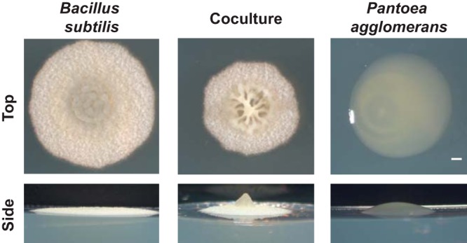
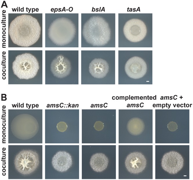
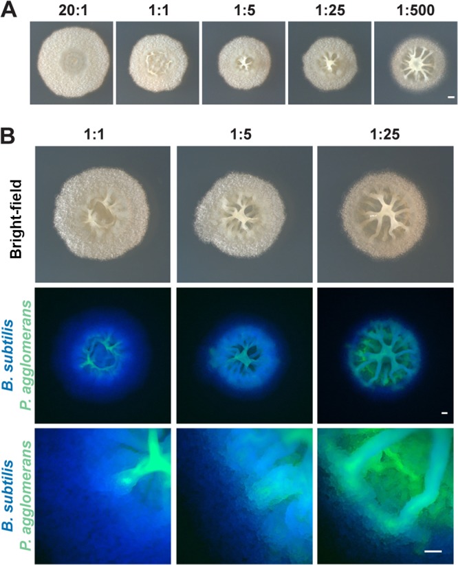
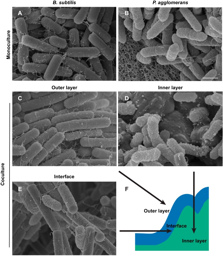
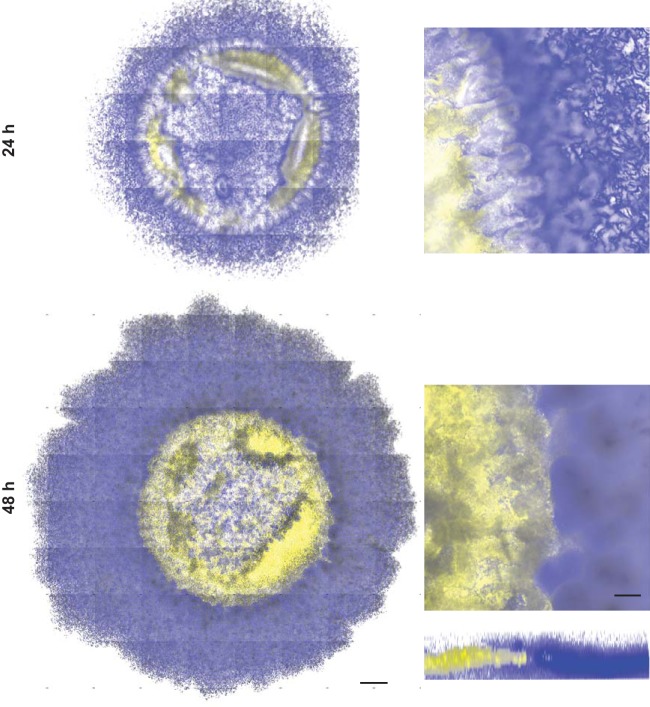
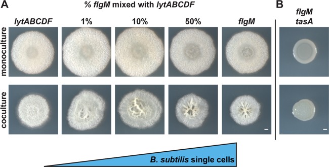
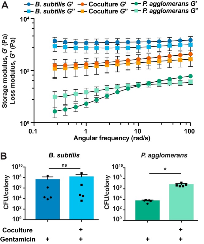
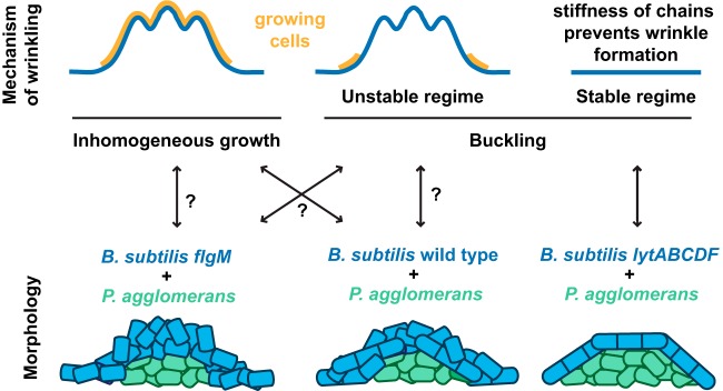
Many microbes coexist within biofilms, or multispecies communities of cells encased in an extracellular matrix. However, little is known about the microbe-microbe interactions relevant for creating these structures. In this study, we explored a striking dual-species biofilm between Bacillus subtilis and Pantoea agglomerans that exhibited characteristics that were not predictable from previous work examining monoculture biofilms. Coculture wrinkle formation required a P. agglomerans exopolysaccharide as well as the B. subtilis amyloid-like protein TasA. Unexpectedly, other B. subtilis matrix components essential for monoculture biofilm formation were not necessary for coculture wrinkling (e.g., the exopolysaccharide EPS, the hydrophobin BslA, and cell chaining). In addition, B. subtilis cell chaining prevented coculture wrinkling, even though chaining was previously associated with more robust monoculture biofilms. We also observed that increasing the relative proportion of P. agglomerans (which forms completely featureless monoculture colonies) increased coculture wrinkling. Using microscopy and rheology, we observed that these two bacteria assemble into an organized layered structure that reflects the physical properties of both monocultures. This partitioning into distinct regions negatively affected the survival of P. agglomerans while also serving as a protective mechanism in the presence of antibiotic stress. Taken together, these data indicate that studying cocultures is a productive avenue to identify novel mechanisms that drive the formation of structured microbial communities.IMPORTANCE In the environment, many microbes form biofilms. However, the interspecies interactions underlying bacterial coexistence within these biofilms remain understudied. Here, we mimic environmentally relevant biofilms by studying a dual-species biofilm formed between Bacillus subtilis and Pantoea agglomerans and subjecting the coculture to chemical and physical stressors that it may experience in the natural world. We determined that both bacteria contribute structural elements to the coculture, which is reflected in its overall viscoelastic behavior. Existence within the coculture can be either beneficial or detrimental depending on the context. Many of the features and determinants of the coculture biofilm appear distinct from those identified in monoculture biofilm studies, highlighting the importance of characterizing multispecies consortia to understand naturally occurring bacterial interactions.

Keywords: Bacillus subtilis; Pantoea agglomerans; antibiotic stress; biofilm; cell-cell interactions; coculture; dual-species; rheology; wrinkles.


Figures

FIG 1
FIG 1
Biofilm architecture of B. subtilis NCIB3610 and environmental soil isolate P. agglomerans in monoculture and coculture grown at 24°C on biofilm-inducing medium MSgg for 48 h. Colony images were taken from the top down to observe wrinkle formation and from the side to visualize the height and 3D structure. Bar, 1 mm.
FIG 2
FIG 2
Extracellular matrix components required for coculture biofilm structure. (A) Colony morphology of B. subtilis strains lacking genes encoding essential matrix components (epsA-O, bslA, and tasA) in monoculture (top) and in coculture with P. agglomerans (bottom) grown on MSgg for 48 h. (B) Representative P. agglomerans mutants (transposon insertion amsC::kan, P. agglomerans amsC, P. agglomerans amsC complemented with amsC on a plasmid [pBBR1MCS-amsC], amsC plus pBBR1MCS [empty vector control]) grown in monoculture (top) and coculture with B. subtilis (bottom) in a 1:500 B. subtilis-to-P. agglomerans ratio on MSgg for 48 h. Bars, 1 mm.
FIG 3
FIG 3
Colony morphology and localization of B. subtilis and P. agglomerans in coculture plated at various initial cell ratios. (A) Colonies of B. subtilis and P. agglomerans in coculture imaged from the top and side at increasing initial ratios of P. agglomerans after growth on MSgg for 48 h. Bar, 1 mm. (B) B. subtilis (false colored blue) and P. agglomerans (false colored green) in coculture at increasing initial ratios of P. agglomerans grown on MSgg after 48 h of growth. Colocalization of two species is indicated in cyan. Coculture images of phase contrast (top) at ×1 magnification, fluorescence (middle) at ×1 magnification, and fluorescence (bottom) at ×8 magnification. Bars, 0.5 mm.
FIG 4
FIG 4
B. subtilis and P. agglomerans cells partition into a layered coculture biofilm. Monoculture and coculture colonies grown on MSgg for 24 h were removed from the agar plate and processed for SEM. (A and B) Cells in B. subtilis monoculture biofilm (A) compared to cells of P. agglomerans in monoculture (B). (C to F) Views of outer (C), inner (D), and interface layers (E) with a schematic of a B. subtilis and P. agglomerans coculture colony (F). Bar, 1 μm.
FIG 5
FIG 5
B. subtilis and P. agglomerans are spatially organized within the coculture biofilm. Coculture colonies grown on glass coverslips embedded in MSgg agar and imaged at 24 h and 48 h using confocal laser scanning microscopy. B. subtilis PspacC-mTurq (blue) and P. agglomerans PspacC-Ypet (yellow) are shown in coculture colony tile scans (left) and an inset of one tile (right) with a representative three-dimensional (3D) reconstructed biofilm shown at 48 h. Bars, 500 μm and 100 μm for tile scans and individual tiles, respectively.
FIG 6
FIG 6
B. subtilis cells in chains prevent formation of the wrinkled coculture phenotype while single cells enhance it. (A) Colonies of B. subtilis lytABCDF (chained cells) cultured with increasing amounts of B. subtilis flgM (single cells) and spotted in monoculture (top) and in coculture with P. agglomerans (bottom) on MSgg and imaged after 48 h of growth. (B) B. subtilis flgM tasA double mutant grown in monoculture (top) and in coculture with P. agglomerans (bottom) on MSgg for 48 h. Bars, 1 mm.
FIG 7
FIG 7
Physical properties of each organism impact the coculture structure and function. (A) Storage modulus (G′, elastic component) and loss modulus (G″, viscous component) of B. subtilis and P. agglomerans biofilms in monoculture and coculture measured over a range of frequencies after 48 h of growth on MSgg (n = 5). Error bars represent standard errors of means. (B) CFU per colony counts of B. subtilis and P. agglomerans in monoculture and coculture biofilms treated with gentamicin for 24 h after 24 h of prior growth. Error bars indicate standard deviations. *, P < 0.05. Statistical significance was determined by a two-tailed t test.
FIG 8
FIG 8
Schematic images illustrating possible mechanisms of wrinkling and the spatial organization of B. subtilis and P. agglomerans within the biofilm. (Top) Inhomogeneous growth occurs when the upper layer of the material grows faster than the interior, leading to wrinkling during growth. Buckling occurs in response to compressive stresses on materials that have already formed. Wrinkles occur only within unstable buckling regimes, when the compressive stresses are above a modulus-dependent threshold; this threshold, along with its wavelengths and amplitude, depends on both the geometry and properties of the thin sheet and its substrate (71, 72). In the stable buckling regime, the material is too stiff for the applied stresses and no buckling occurs. (Bottom) The colony morphology of each coculture is shown with the B. subtilis strain in blue (forming an elastic shell) and the P. agglomerans strain in green (forming a central “droplet” of more viscous material).

Similar articles

Cited by

References

    1. Hall-Stoodley L, Costerton JW, Stoodley P. 2004. Bacterial biofilms: from the natural environment to infectious diseases. Nat Rev Microbiol 2:95–108. doi:10.1038/nrmicro821. - DOI - PubMed
    1. Flemming H-C, Wingender J. 2010. The biofilm matrix. Nat Rev Microbiol 8:623–633. doi:10.1038/nrmicro2415. - DOI - PubMed
    1. Mah TF, O'Toole GA. 2001. Mechanisms of biofilm resistance to antimicrobial agents. Trends Microbiol 9:34–39. doi:10.1016/S0966-842X(00)01913-2. - DOI - PubMed
    1. Hobley L, Harkins C, MacPhee CE, Stanley-Wall NR. 2015. Giving structure to the biofilm matrix: an overview of individual strategies and emerging common themes. FEMS Microbiol Rev 39:649–669. doi:10.1093/femsre/fuv015. - DOI - PMC - PubMed
    1. Matz C, Kjelleberg S. 2005. Off the hook–how bacteria survive protozoan grazing. Trends Microbiol 13:302–307. doi:10.1016/j.tim.2005.05.009. - DOI - PubMed

Publication types

MeSH terms

Substances

LinkOut - more resources