. 2019 Mar 19;116(12):5785-5794.
doi: 10.1073/pnas.1821000116. Epub 2019 Mar 4.

AAV cis-regulatory sequences are correlated with ocular toxicity

Affiliations

AAV cis-regulatory sequences are correlated with ocular toxicity

Wenjun Xiong et al. Proc Natl Acad Sci U S A. .

Abstract

Adeno-associated viral vectors (AAVs) have become popular for gene therapy, given their many advantages, including their reduced inflammatory profile compared with that of other viruses. However, even in areas of immune privilege such as the eye, AAV vectors are capable of eliciting host-cell responses. To investigate the effects of such responses on several ocular cell types, we tested multiple AAV genome structures and capsid types using subretinal injections in mice. Assays of morphology, inflammation, and physiology were performed. Pathological effects on photoreceptors and the retinal pigment epithelium (RPE) were observed. Müller glia and microglia were activated, and the proinflammatory cytokines TNF-α and IL-1β were up-regulated. There was a strong correlation between cis-regulatory sequences and toxicity. AAVs with any one of three broadly active promoters, or an RPE-specific promoter, were toxic, while AAVs with four different photoreceptor-specific promoters were not toxic at the highest doses tested. There was little correlation between toxicity and transgene, capsid type, preparation method, or cellular contaminants within a preparation. The toxic effect was dose-dependent, with the RPE being more sensitive than photoreceptors. Our results suggest that ocular AAV toxicity is associated with certain AAV cis-regulatory sequences and/or their activity and that retinal damage occurs due to responses by the RPE and/or microglia. By applying multiple, sensitive assays of toxicity, AAV vectors can be designed so that they can be used safely at high dose, potentially providing greater therapeutic efficacy.

Keywords: AAV; gene therapy; retina; retinal pigment epithelium; toxicity.


Conflict of interest statement

Conflict of interest statement: The authors note we have received funding from Astellas for our work.

Figures
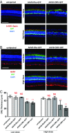
Fig. 1.
Fig. 1.
Retina toxicity is induced by broadly active but not retinal cell-type-specific promoters. (A and B) Wild-type retinas of CD-1 mice were infected at P0 with the indicated viruses at either high (3E9 vg per eye) or low dose (8E8 vg per eye) and harvested at P30 for histology. Retinal cross-sections were stained for short- and medium/long-wavelength opsins (red) (A) and for GFAP (red) (B). Loss of opsin staining and up-regulated expression of GFAP were observed in the retinas infected with AAV8-CMV-GFP. (Scale bars, 100 µm.) (C) Quantification of ONL thickness at 1 mm from optic nerve head (ONH). Data are presented as mean ± SD. n = 3–17 per group. One-way ANOVA analysis with Tukey test, **P < 0.01; NS, not significant between the designated group and the uninjected group.
Fig. 2.
Fig. 2.
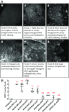
RPE toxicity is induced by promoters that are active in the RPE. (A) Grading criteria of RPE toxicity, with grade 0 representing completely healthy RPE and grade 5 representing the most severe RPE damage. The typical phenotypes of each grade are described below each image. (Scale bars, 50 μm.) (B) Scatter dot plot of RPE toxicity grades. All viruses were tested at a dose of 8E8 vg per eye, except for AAV8-CAR-GFP (3E9 vg per eye), in CD-1 mice injected at P0 and killed at P30. Data are presented as mean ± SD. n = 2–8 per group. One-way ANOVA analysis with Tukey test, **P < 0.01; NS, not significant between the designated group and the uninjected group.
Fig. 3.
Fig. 3.
RPE and retina damage is dose-dependent. Representative images of RPE and retina of P30 CD-1 mice following infection at P0 with low dose (5E8 vg per eye), medium dose (1E9 vg per eye) and high dose (2E9 vg per eye) of AAV8-CMV-GFP. (AC) The RPE (labeled with phalloidin staining) and (DF) photoreceptors (labeled with PNA) from the same areas are shown in upper and lower panels. (Scale bars, 50 µm.)
Fig. 4.
Fig. 4.
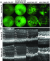
OCT images of eyes infected by AAV8-CMV-GFP and AAV5-CMV-GFP. (A) Funduscopic images of eyes from C57BL/6J mice infected at P0 with low (L: 8E8 vg per eye) and high (H: 3E9 vg per eye) doses of AAV8-CAR-GFP, AAV8-RedO-GFP + AAV8-Best1-GFP (5:1 ratio), AAV8-CMV-GFP, and AAV5-CMV-GFP viruses. Infected mouse eyes were examined at approximately P30. Green arrow labels the plane where the OCT image was taken. (B) OCT images of the eyes shown in A. White arrowhead, intrusions in subretinal space. BV, blood vessel; INL, inner nuclear layer; IPL, inner plexiform layer; IS/OS, junction of inner and outer segments of photoreceptors; OPL, outer plexiform layer.
Fig. 5.
Fig. 5.
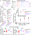
Toxic AAV causes ERG and optomotor manifestations in C57BL/6J mouse eyes. (A) Representative trace of scotopic ERG (flash intensity: 0.1 cd s/m2, wavelength: 530 nm) of P30 eyes injected at P0 with AAV8-RedO-GFP + AAV8-Best1-GFP (5:1 ratio, RedO+Best1) or AAV8-CMV-GFP (CMV) at low dose or high dose. (B) Statistics of scotopic ERG parameters (a-wave and b-wave amplitude and implicit time) of P30 eyes injected at P0 with RedO+Best1 (low dose: n = 8, high dose: n = 7) and CMV (low dose: n = 10, high dose: n = 8). (C) Representative traces of photopic ERG of P30 eyes injected at P0 with RedO+Best1 and CMV, elicited by 1 (peak), 10 (peak), 100 (xenon), and 1,000 (xenon) cd s/m2 white light flashes with a white light background (bkg) of 30 cd/m2. (D) Ensemble-averaged photopic ERG b-wave amplitude from P30 eyes injected at P0 with RedO+Best1 (low dose: n = 8, high dose: n = 7) and CMV (low dose: n = 10, high dose: n = 8). Significance (P value): CMV (H) vs. RedO+Best1 (H) at 1 (****), 10 (*#), 100 (****), and 1,000 (****) cd s/m2; CMV (L) vs. RedO+Best1 (L) at 1 (**), 10 (*), 100 (NS), and 1,000 (NS) cd s/m2. (Inset) The normalized photopic ERG b-wave intensity–response (r/rmax) curves with I1/2 of RedO+Best1 (low dose: 4.8 cd s/m2, high dose: 5.8 cd s/m2), and CMV (low dose: 9.0 cd s/m2, high dose: 27.0 cd s/m2). (E) Photopic optomotor responses from approximately P35 eyes injected either with RedO+Best1 (low dose: n = 7, high dose: n = 7) or CMV (low dose: n = 13, high dose: n = 7). Acuity: tested at 100% contrast; contrast sensitivity: tested at 0.128 cyc/deg and 1.5-Hz temporal frequency. For all panels, data are presented as mean ± SEM, unpaired Student’s t test *P < 0.05, ***P < 0.001, ****P < 0.0001, *#P < 1 × 10⁻⁶, ***#P < 1 × 10⁻⁸; NS, not significant. CMV, AAV8-CMV-GFP; H, high dose; L, low dose; RedO+Best1 for AAV8-RedO-GFP +AAV8-Best1-GFP (5:1 ratio).
Fig. 6.
Fig. 6.
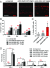
Activation of microglia and innate immune response by toxic AAVs. (A) Iba1 staining of retinal sections from P30 eyes of CD-1 mice infected at P0 with the indicated AAV viruses at a dose of 3E9 vg per eye. (Scale bar: 50 μm.) (B) Quantification of displaced Iba1-positive cells by cell layer. High dose (3E9 vg per eye); low dose (8E8 vg per eye). Values are shown as mean ± SD. n = 4 per group. (C) Quantification of microglia in retinas of P20 Cx3cr1-GFP mice by flow cytometry injected with PBS or 3E8 vg per eye AAV8-CMV-TdTomato (n = 3–4 for all groups). Values are shown as mean ± SD. (D) Relative mRNA levels of TNF-α, IL-1β, IL-6, and IFN-γ by qPCR in the retinas of P30 CD-1 mice infected at P0 with the indicated AAV viruses at high dose (3E9 vg per eye). Expression level was normalized to gapdh. Values are shown as mean ± SEM. n = 4–8 per group. One-way ANOVA analysis with Tukey test, **P < 0.01; *P < 0.05; NS, not significant between the designated group and the uninjected group.

References

    1. Gao G, et al. Clades of adeno-associated viruses are widely disseminated in human tissues. J Virol. 2004;78:6381–6388. - PMC - PubMed
    1. Dalkara D, et al. In vivo-directed evolution of a new adeno-associated virus for therapeutic outer retinal gene delivery from the vitreous. Sci Transl Med. 2013;5:189ra76. - PubMed
    1. Zinn E, et al. In silico reconstruction of the viral evolutionary lineage yields a potent gene therapy vector. Cell Rep. 2015;12:1056–1068. - PMC - PubMed
    1. Bainbridge JWB, et al. Effect of gene therapy on visual function in Leber’s congenital amaurosis. N Engl J Med. 2008;358:2231–2239. - PubMed
    1. Hauswirth WW, et al. Treatment of Leber congenital amaurosis due to RPE65 mutations by ocular subretinal injection of adeno-associated virus gene vector: Short-term results of a phase I trial. Hum Gene Ther. 2008;19:979–990. - PMC - PubMed

Publication types