Review
. 2020 Jan 2;12(1):a033985.
doi: 10.1101/cshperspect.a033985.

The Proteasome and Its Network: Engineering for Adaptability

Affiliations
Review

The Proteasome and Its Network: Engineering for Adaptability

Daniel Finley et al. Cold Spring Harb Perspect Biol. .

Abstract

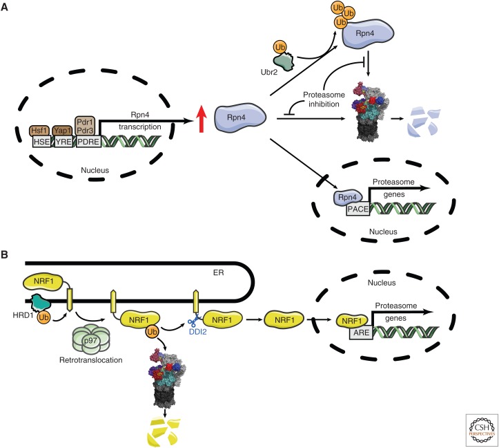
The proteasome, the most complex protease known, degrades proteins that have been conjugated to ubiquitin. It faces the unique challenge of acting enzymatically on hundreds and perhaps thousands of structurally diverse substrates, mechanically unfolding them from their native state and translocating them vectorially from one specialized compartment of the enzyme to another. Moreover, substrates are modified by ubiquitin in myriad configurations of chains. The many unusual design features of the proteasome may have evolved in part to endow this enzyme with a robust ability to process substrates regardless of their identity. The proteasome plays a major role in preserving protein homeostasis in the cell, which requires adaptation to a wide variety of stress conditions. Modulation of proteasome function is achieved through a large network of proteins that interact with it dynamically, modify it enzymatically, or fine-tune its levels. The resulting adaptability of the proteasome, which is unique among proteases, enables cells to control the output of the ubiquitin-proteasome pathway on a global scale.


Figures

Figure 1.
Figure 1.
Conformational states of substrate-bound human proteasomes. Cryo-electron microscopy (cryo-EM) density maps of substrate (orange)-bound human proteasomes in three different stages of substrate processing are represented. (A) A conformation that is compatible with the deubiquitylation of the bound substrate. (B,C) Two consecutive conformations of substrate translocation (EC1 and ED2) into the core particle (CP). Color coding for the proteasome lid, the RPT/ATPase ring, and CP is given in the legend to Fig. 2. RPT1 and RPT5 density maps were removed from the ATPase rings to expose the substrate translocation channel of the regulatory particle (RP). In ED2, subunit α1 (PSMA1) was also removed to show the substrate translocation channel of the CP. EB (Protein Data Bank [PDB]: 6MSE), EC1 (PDB: 6MSG), and ED2 (PDB: 6MSK) density maps were obtained from data in Dong et al. (2018).
Figure 2.
Figure 2.
Overview of deubiquitinating enzymes (DUBs) and Ub/Ubl receptors of the human proteasome. A cryo-electron microscopy (cryo-EM) density map of the human proteasome is represented with all of its known ubiquitin receptors (RPN1, RPN10, and RPN13 in purple tones) and DUBs (RPN11, USP14, and UCH37 in red tones). The lid subassembly of the RP is in light gray. The cryo-EM structure of the proteasome bound to USP14-ubiquitin-aldehyde was obtained from data presented in Huang et al. (2016b) (Protein Data Bank [PDB]: 5GJQ). The location of UCH37 and RPN13 were modeled as shown in de Poot et al. (2017) using data in density maps from Sahtoe et al. (2015) and Vander Linden et al. (2015) (PDB: 4UEL and 4WLQ). All six subunits of the RPT ring are shown in light blue and ubiquitin monomers bound to the DUBs are shown in yellow ribbon representations.
Figure 3.
Figure 3.
Extrinsic ubiquitin receptors of the proteasome. Shown is a schematic representation of four protein families of Saccharomyces cerevisiae and Homo sapiens, which include proteins thought to deliver ubiquitinated targets to the proteasome (Rad23, Ddi1, Dsk2, and Zfand). Some of these proteins do not appear to be functional ubiquitin receptors (see text for details). The simple modular architecture research tool (SMART) (Letunic and Bork 2018) was used to identify SMART and Pfam protein domains. All proteins and domains are drawn to scale.
Figure 4.
Figure 4.
Representation of the ATP hydrolytic cycle and substrate translocation. (A,B) Two different cryo-electron microscopy (cryo-EM) structures of the RPT ring at different states of substrate (orange mesh) translocation in the yeast proteasome holoenzyme are shown (states 5D and 4D, respectively). Images were created from data in de la Pena et al. (2018). Ribbon representations of the top view of the RPT rings are shown in the bottom panels, where substrate disengagement of Rpt5 (green) and Rpt4 (blue) can be observed (A and B, respectively). A 90° rotation about the x axis was applied to show a lateral view of the RPT ring (top panels). While the substrate polypeptide is surrounded by a spiral-staircase of pore 1 loop tyrosines (highlighted in pink) projecting from substrate-engaged RPT subunits, the Tyr of the RPT subunits that are disengaged (Rpt5 in A and Rpt4 in B) are displaced from the substrate. In the bottom panels, spherical representations of ATP (yellow) and ADP (blue) are also included. For a better view of the spiral-staircase model in the top panels, two RPT subunits (Rpt4 and Rpt3) and three amino acids (Arg261, Glu293, and Gly294 of RPT4) were omitted from the density maps. States 5D and 4D were created from data in de la Pena et al. (2018) (Protein Data Bank [PDB]: 6EF1 and 6EF3, respectively).
Figure 5.
Figure 5.
Feedback control of proteasome levels in yeast and mammals. (A) In S. cerevisiae, transcription factor Rpn4 controls the level of expression of proteasome subunits. Rpn4 is stabilized under specific proteotoxic stresses, inducing the transcription of proteasomal subunit genes regulated by proteasome-associated control (PACE) elements. (B) In mammals, NRF1 is the key transcriptional regulator of proteasome subunit genes. A resident of the endoplasmic reticulum (ER), NRF1 is ubiquitinated by HRD1 and retrotranslocated by p97. NRF1 protein can then be degraded via the proteasome as an ERAD substrate or cleaved endoproteolytically by the protease DDI2, resulting in its liberation from the ER membrane. The cleaved version of NRF1 is translocated to the nucleus where it binds ARE elements and induces the transcription of genes for proteasome subunits. The nuclear form of NRF1 is also rapidly degraded by the proteasome (see main text).
Figure 6.
Figure 6.
Activation and inhibition of USP14. (A) Structure of the substrate-engaged USP14 (red) bound to ubiquitin aldehyde (Ub-Al, in yellow), a model of the enzyme's activated state. The principal difference from the ubiquitin-free (inactive) state of USP14 is in the positioning of the two blocking (BL) loops (Hu et al. 2005). The BL loops of USP14-Ub-Al are in green, those of the free USP14 in blue. To the right of these loops, the active-site Cys is represented as a light green patch. In the free form of USP14, the BL loops block the access of the carboxyl terminus of ubiquitin to the active site. This occlusion is relieved in the substrate-engaged form. Ser432 (gray), located within the BL2 loop, is subject to phosphorylation by AKT, providing partial activation (Xu et al. 2015). (B) Structure of USP14 bound to IU1-47 (white) shows that this inhibitor occludes access of the carboxyl terminus of ubiquitin to the catalytic site (Wang et al. 2018). To better visualize the IU1-47-binding pocket, both BL loops were removed from the density maps. (C) Structure of free USP14 with IU1-47 superimposed. This model shows how the BL2 loop (blue) occludes access of IU1-47, consistent with previous evidence that IU1 series compounds do not inhibit the free form of USP14 (Lee et al. 2016). Density maps of the USP14 catalytic domain in its free form (Protein Data Bank [PDB]: 2AYN), USP14 bound to Ub-Al (PDB: 2AYO), and the USP14-IU1-47 complex (PDB: 6IIL) were created from data in Hu et al. (2005) and Wang et al. (2018). To better visualize the catalytic site of USP14, Gln197 was removed from all USP14 structures in this figure.

References

    1. Asano S, Fukuda Y, Beck F, Aufderheide A, Forster F, Danev R, Baumeister W. 2015. Proteasomes. A molecular census of 26S proteasomes in intact neurons. Science 347: 439–442. 10.1126/science.1261197 - DOI - PubMed
    1. Aufderheide A, Beck F, Stengel F, Hartwig M, Schweitzer A, Pfeifer G, Goldberg AL, Sakata E, Baumeister W, Förster F. 2015. Structural characterization of the interaction of Ubp6 with the 26S proteasome. Proc Natl Acad Sci 112: 8626–8631. 10.1073/pnas.1510449112 - DOI - PMC - PubMed
    1. Aviram S, Kornitzer D. 2010. The ubiquitin ligase Hul5 promotes proteasomal processivity. Mol Cell Biol 30: 985–994. 10.1128/MCB.00909-09 - DOI - PMC - PubMed
    1. Bajorek M, Finley D, Glickman MH. 2003. Proteasome disassembly and downregulation is correlated with viability during stationary phase. Curr Biol 13: 1140–1144. 10.1016/S0960-9822(03)00417-2 - DOI - PubMed
    1. Bard JAM, Goodall EA, Greene ER, Jonsson E, Dong KC, Martin A. 2018. Structure and function of the 26S proteasome. Annu Rev Biochem 87: 697–724. 10.1146/annurev-biochem-062917-011931 - DOI - PMC - PubMed

Publication types

MeSH terms

LinkOut - more resources