Review
. 2019 Nov 1;11(11):a033902.
doi: 10.1101/cshperspect.a033902.

Recognition and Degradation of Mislocalized Proteins in Health and Disease

Affiliations
Review

Recognition and Degradation of Mislocalized Proteins in Health and Disease

Ramanujan S Hegde et al. Cold Spring Harb Perspect Biol. .

Abstract

A defining feature of eukaryotic cells is the segregation of complex biochemical processes among different intracellular compartments. The protein targeting, translocation, and trafficking pathways that sustain compartmentalization must recognize a diverse range of clients via degenerate signals. This recognition is imperfect, resulting in polypeptides at incorrect cellular locations. Cells have evolved mechanisms to selectively recognize mislocalized proteins and triage them for degradation or rescue. These spatial quality control pathways maintain cellular protein homeostasis, become especially important during organelle stress, and might contribute to disease when they are impaired or overwhelmed.


Figures

Figure 1.
Figure 1.
The recognition problem for hydrophobic targeting sequences. Shown are examples of an endoplasmic reticulum (ER)-destined signal sequence (only the hydrophobic domain is depicted), the first transmembrane domains for proteins destined for the ER (opioid receptor) and mitochondria (ADP/ATP carrier), and a mitochondrial presequence (only the amphipathic helix is depicted). Despite many similarities among the four targeting sequences, the first two need to be recognized by the M-domain of SRP54, whereas the other two need to be avoided. The amino acids in the targeting sequences are colored as follows: acidic in pink, basic in light blue, highly hydrophobic in dark green, moderately hydrophobic in pale green, and polar in yellow. The substrate binding M-domain of SRP54 is taken from Protein Data Bank (PDB) code 3JAN, which was modeled from 1QB2, the X-ray structure of the human SRP54 M-domain (Clemons et al. 1999). Hydrophobic residues of SRP54 are colored green.
Figure 2.
Figure 2.
Multiple fates for an endoplasmic reticulum (ER)-destined mislocalized protein. Protein import into the ER can fail, resulting in a mislocalized protein in the cytosol (inside the red oval in the center). This failure occurs constitutively at a low level under normal conditions (red arrows, left side) but can be enhanced for some proteins during acute ER stress (red arrows, right side). The mislocalized protein can be degraded, aggregate with itself or other proteins, or mistarget to another organelle.
Figure 3.
Figure 3.
Protein mislocalization and signaling during mito. (mitochondrial) stress. Depicted are the fates of several mitochondrial precursors during normal conditions (left) and acute stress (right). Failed import during acute stress results in (presumably misfolded) mitochondrial precursors in the cytosol that can trigger proteasomal up-regulation by a mechanism that is incompletely understood (Wrobel et al. 2015). In special cases, the nonimported precursor is not degraded selectively during mitochondrial stress and can signal downstream responses. This is the case for ATFS1, which can act as a transcription factor to up-regulate genes that improve mitochondrial homeostasis (Nargund et al. 2012). PINK1 is normally imported into the inner membrane where its cleavage permits retrotranslocation to the cytosol for degradation. During acute stress, PINK1 import to the inner membrane is aborted, allowing its insertion into the mitochondrial outer membrane where it initiates a series of events leading to mitochondrial autophagy (Sekine and Youle 2018). Pdr3 is also activated during certain types of mitochondrial stress (Weidberg and Amon 2018), although the signal for activation, perhaps a mitochondrial metabolite, is not known.
Figure 4.
Figure 4.
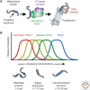
Cytosolic recognition of mislocalized proteins. (A) Targeting signals (or transmembrane domains) can be recognized by dedicated cytosolic factors that recruit ubiquitination machinery to mediate substrate ubiquitination. These substrates are then degraded by proteasomes. (B) Schematic depiction of the concept that different cytosolic factors, all of which recognize exposed hydrophobicity, are tuned to different lengths and degrees of hydrophobic domains. Biochemical analyses suggest that there is substantial overlap in what is recognized.
Figure 5.
Figure 5.
Recognition of mistargeted proteins at organelle membranes. Mistargeted proteins are depicted at the endoplasmic reticulum (ER) and mitochondrion. At the ER, they might be recognized by E3 ubiquitin ligase complexes centered around TRC8 or MARCH6 (Doa10 in yeast) and routed for degradation in the cytosol (Metzger et al. 2008; Stefanovic-Barrett et al. 2018). At mitochondria, the AAA+ ATPase Msp1 (ATAD1 in mammals) can recognize mistargeted proteins, possibly with the aid of an adaptor, to mediate extraction into the cytosol (Chen et al. 2014; Okreglak and Walter 2014; Weir et al. 2017; Wohlever et al. 2017; Weidberg and Amon 2018). Whether ubiquitin ligases are involved and at what step they act to mediate Msp1-mediated degradation is not known. Mistargeted proteins can also be rescued, as exemplified by a pathway from the ER to mitochondria involving the chaperone Djp1 (Hansen et al. 2018). Misloc. protein, Mislocalized protein.

References

    1. Arnold A, Horst SA, Gardella TJ, Baba H, Levine MA, Kronenberg HM. 1990. Mutation of the signal peptide-encoding region of the preproparathyroid hormone gene in familial isolated hypoparathyroidism. J Clin Invest 86: 1084–1087. 10.1172/JCI114811 - DOI - PMC - PubMed
    1. Aviram N, Ast T, Costa EA, Arakel EC, Chuartzman SG, Jan CH, Haßdenteufel S, Dudek J, Jung M, Schorr S, et al. 2016. The SND proteins constitute an alternative targeting route to the endoplasmic reticulum. Nature 540: 134–138. 10.1038/nature20169 - DOI - PMC - PubMed
    1. Borgese N, Fasana E. 2011. Targeting pathways of C-tail-anchored proteins. Biochim Biophys Acta 1808: 937–946. 10.1016/j.bbamem.2010.07.010 - DOI - PubMed
    1. Brandman O, Stewart-Ornstein J, Wong D, Larson A, Williams CC, Li GW, Zhou S, King D, Shen PS, Weibezahn J, et al. 2012. A ribosome-bound quality control complex triggers degradation of nascent peptides and signals translation stress. Cell 151: 1042–1054. 10.1016/j.cell.2012.10.044 - DOI - PMC - PubMed
    1. Büeler H, Aguzzi A, Sailer A, Greiner RA, Autenried P, Aguet M, Weissmann C. 1993. Mice devoid of PrP are resistant to scrapie. Cell 73: 1339–1347. 10.1016/0092-8674(93)90360-3 - DOI - PubMed

Publication types

LinkOut - more resources