Review
. 2020 Jan 2;12(1):a033951.
doi: 10.1101/cshperspect.a033951.

Functional Modules of the Proteostasis Network

Affiliations
Review

Functional Modules of the Proteostasis Network

Gopal G Jayaraj et al. Cold Spring Harb Perspect Biol. .

Abstract

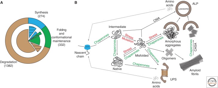
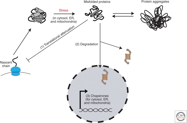
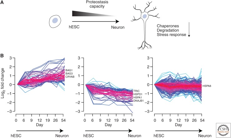
Cells invest in an extensive network of factors to maintain protein homeostasis (proteostasis) and prevent the accumulation of potentially toxic protein aggregates. This proteostasis network (PN) comprises the machineries for the biogenesis, folding, conformational maintenance, and degradation of proteins with molecular chaperones as central coordinators. Here, we review recent progress in understanding the modular architecture of the PN in mammalian cells and how it is modified during cell differentiation. We discuss the capacity and limitations of the PN in maintaining proteome integrity in the face of proteotoxic stresses, such as aggregate formation in neurodegenerative diseases. Finally, we outline various pharmacological interventions to ameliorate proteostasis imbalance.


Figures

Figure 1.
Figure 1.
Architecture of the proteostasis network (PN). (A) The three modules of the PN (outer ring): synthesis (blue, 274 factors), folding and conformational maintenance (green, 332 factors), and degradation (brown, 1382 factors). The inner pie chart shows the partitioning of each module into essential (hatched sectors, synthesis [203/274], folding [78/332], and degradation [178/1382]) and nonessential components (filled sectors). (B) Central role of molecular chaperones in the PN. The nascent polypeptide chain emerging from the ribosomal tunnel is prevented from misfolding and aggregation by chaperones, including components that bind to the ribosome (trigger factor in bacteria, nascent chain–associated complex [NAC] and ribosome-associated complex [RAC] in eukarya). A second tier of chaperones (Hsp70, Hsp90, chaperonins) does not interact with the ribosome directly and mediates co- or posttranslational folding. Proteins that misfold because of mutations or under conditions of stress are selectively degraded either by the ubiquitin–proteasome system (UPS) or chaperone-mediated autophagy (CMA) and chaperone-assisted selective autophagy (CASA). Misfolded proteins may aggregate to soluble oligomers, amorphous aggregates, or amyloid fibrils when basal chaperone and degradation capacity is exceeded. Aggregates may be sequestered into insoluble inclusions, which may be dissociated into fragments by specialized chaperones (Hsp104 in yeast, Hsp70/Hsp40/Hsp110 in metazoans) for subsequent clearance by the autophagy–lysosomal pathway (ALP).
Figure 2.
Figure 2.
Proteostasis capacity and stress responses. During proteotoxic stress in different cellular compartments, proteostasis capacity is adjusted to meet increased cellular requirements. Global translational attenuation (1) reduces the burden on the folding machineries to free chaperone capacities for assistance in clearance of misfolded and aggregated species (2). Concomitantly, transcriptional stress response pathways are activated (3) to increase chaperone pools available for folding and degradation. ER, Endoplasmic reticulum.
Figure 3.
Figure 3.
Proteostasis network (PN) rewiring and decline in proteostasis capacity during cell differentiation. (A) Human embryonic stem cells (hESCs) have a higher proteostasis capacity as compared to differentiated cells, such as neurons. As hESCs differentiate to neurons, the chaperonins (cytosolic TRiC/CCT and mitochondrial Hsp60) decrease in abundance and there is a general decline in degradation capacity. Additionally, differentiated neurons are less efficient in mounting a stress response in comparison to their progenitors and stem cells. (B) Chaperome trajectories during neuronal differentiation of hESCs (332 proteins analyzed). The chaperome landscape changes in abundance (indicated as log2 fold change) as hESCs are differentiated into neurons. Chaperones can increase (left), decrease (middle), or remain unchanged in abundance (right) during this process. Prominent examples of each cluster are shown (BAG1/BAG3, regulators of Hsp70/Hsc70; CRYAB, α-crystallin B chain; TRiC, cytosolic chaperonin; HSPD1, mitochondrial Hsp60; HSPA1, Hsp70; DNAJB1, Hsp40 regulator of Hsp70/Hsc70; HSPA8, Hsc70). Data for time-dependent changes in chaperone abundance values were derived from Sequence Read Archive (SRA) accession No. GSE86985 (Yao et al. 2017). TPM (transcripts per million) values were directly used to calculate clustering of chaperone trajectories using the fuzzy c-means algorithm Mfuzz (Kumar and Futschik 2007) with “m” values set at 2 and a minimum membership cutoff of 0.8. The gradient from red to blue indicates high to low membership scores.

References

    1. Albanèse V, Yam AY, Baughman J, Parnot C, Frydman J. 2006. Systems analyses reveal two chaperone networks with distinct functions in eukaryotic cells. Cell 124: 75–88. 10.1016/j.cell.2005.11.039 - DOI - PubMed
    1. Anckar J, Sistonen L. 2011. Regulation of HSF1 function in the heat stress response: Implications in aging and disease. Annu Rev Biochem 80: 1089–1115. 10.1146/annurev-biochem-060809-095203 - DOI - PubMed
    1. Anfinsen CB. 1973. Principles that govern the folding of protein chains. Science 181: 223–230. 10.1126/science.181.4096.223 - DOI - PubMed
    1. Arndt V, Dick N, Tawo R, Dreiseidler M, Wenzel D, Hesse M, Fürst DO, Saftig P, Saint R, Fleischmann BK, et al. 2010. Chaperone-assisted selective autophagy is essential for muscle maintenance. Curr Biol 20: 143–148. 10.1016/j.cub.2009.11.022 - DOI - PubMed
    1. Balch WE, Morimoto RI, Dillin A, Kelly JW. 2008. Adapting proteostasis for disease intervention. Science 319: 916–919. 10.1126/science.1141448 - DOI - PubMed

Publication types