. 2019 May;68(5):975-987.
doi: 10.2337/db18-0761. Epub 2019 Mar 4.

DNA Damage Does Not Cause BrdU Labeling of Mouse or Human β-Cells

Affiliations

DNA Damage Does Not Cause BrdU Labeling of Mouse or Human β-Cells

Rohit B Sharma et al. Diabetes. 2019 May.

Abstract

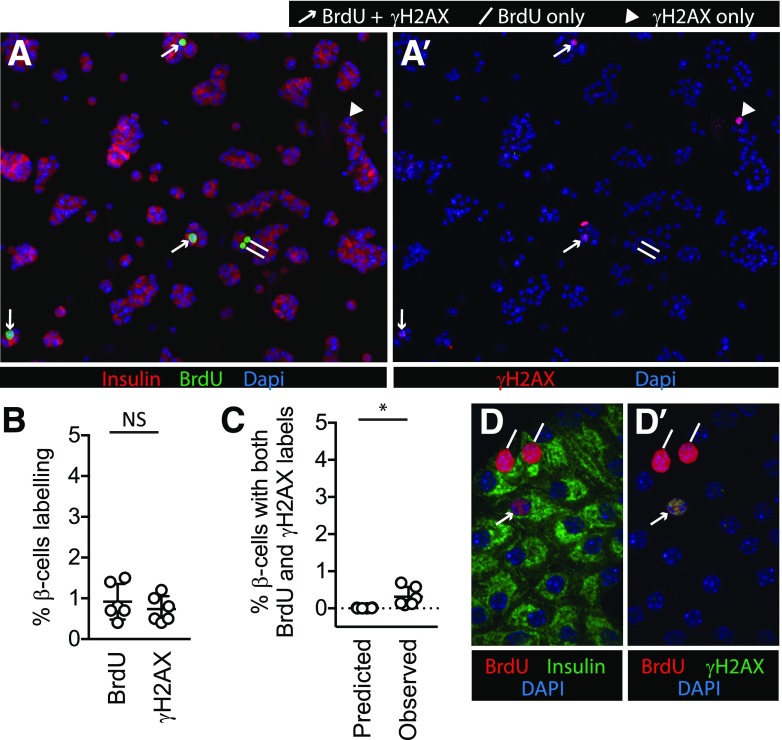
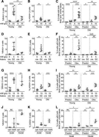
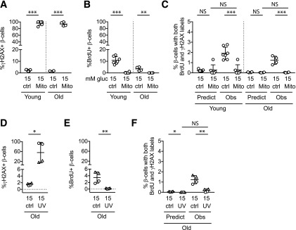
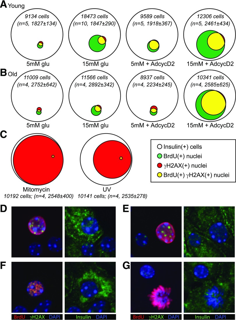
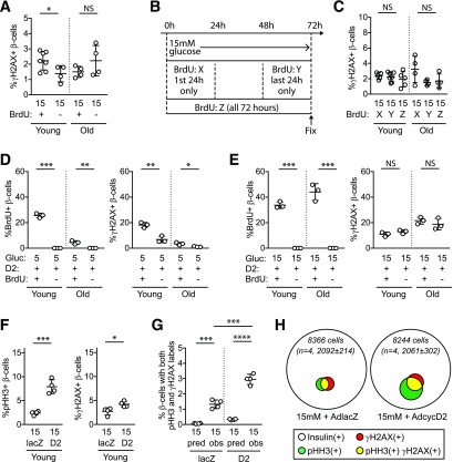
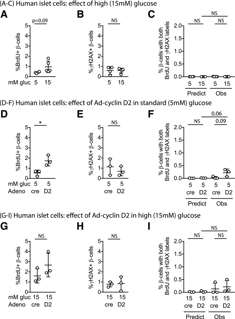
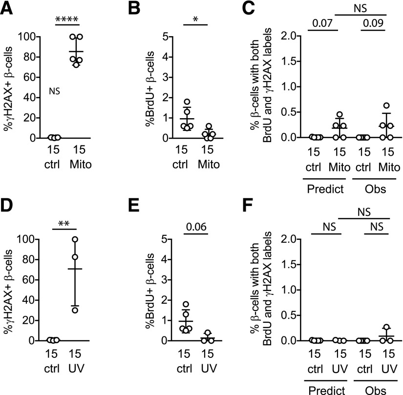
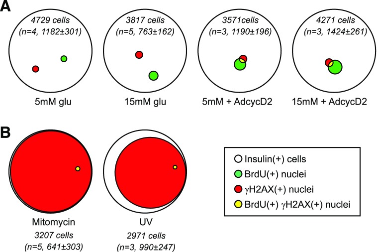
Pancreatic β-cell regeneration, the therapeutic expansion of β-cell number to reverse diabetes, is an important goal. Replication of differentiated insulin-producing cells is the major source of new β-cells in adult mice and juvenile humans. Nucleoside analogs such as BrdU, which are incorporated into DNA during S-phase, have been widely used to quantify β-cell proliferation. However, reports of β-cell nuclei labeling with both BrdU and γ-phosphorylated H2A histone family member X (γH2AX), a DNA damage marker, have raised questions about the fidelity of BrdU to label S-phase, especially during conditions when DNA damage is present. We performed experiments to clarify the causes of BrdU-γH2AX double labeling in mouse and human β-cells. BrdU-γH2AX colabeling is neither an age-related phenomenon nor limited to human β-cells. DNA damage suppressed BrdU labeling and BrdU-γH2AX colabeling. In dispersed islet cells, but not in intact islets or in vivo, pro-proliferative conditions promoted both BrdU and γH2AX labeling, which could indicate DNA damage, DNA replication stress, or cell cycle-related intrinsic H2AX phosphorylation. Strategies to increase β-cell number must not only tackle the difficult challenge of enticing a quiescent cell to enter the cell cycle, but also achieve safe completion of the cell division process.


Figures

Figure 1
Figure 1
BrdU and γH2AX labeling co-occur in some β-cells in mouse islet cell cultures. Dispersed mouse islet cells were cultured for 72 h in 5 mmol/L glucose (A, A′, B, and C) or 15 mmol/L glucose (D and D′), with BrdU added for the final 24 h. BrdU (A) and γH2AX (A′) each labeled a small fraction of β-cells under unstimulated culture conditions. In mouse islet cells cultured in unstimulated (5 mmol/L glucose) conditions, a small proportion of β-cells were labeled with BrdU or γH2AX (B). The observed fraction of β-cells colabeled with both BrdU and γH2AX was low, but higher than that predicted by random chance (the predicted fraction was defined by the product of the observed fractions of each single label) (C). D and D′: Confocal microscopy of mouse islet cells cultured in 15 mmol/L glucose confirmed both labels occur in the same nuclei. For A, A′, D, and D, lines mark BrdU(+) γH2AX(−) nuclei, arrowheads mark BrdU(−) γH2AX(+) nuclei, and arrows mark BrdU(+) γH2AX(+) nuclei. *P < 0.05; NS, P > 0.1.
Figure 2
Figure 2
Proliferative stimuli, especially in combination, increase BrdU-γH2AX colocalization frequency in mouse β-cells. Dispersed young (10- to 12-week-old) and old (50- to 60-week-old) mouse islet cells were cultured for 72 h in the indicated conditions, with BrdU added for the final 24 h. A 15 mmol/L glucose concentration markedly increased BrdU incorporation (A), especially in young islets, and modestly increased γH2AX labeling (B) and BrdU-γH2AX colabeling (C). Ad-cyclin D2 in 5 mmol/L glucose increased BrdU (D), γH2AX (E), and colabeled cells (F). Combined treatment with 15 mmol/L glucose and Ad-cyclin D2 markedly increased BrdU (G), γH2AX (H), and colabeled cells (I). Exposure to a different β-cell mitogen, harmine, also increased BrdU (J), γH2AX (K), and colabeled cells (L). Note the variable y-axis scale in AL. C, F, I, and L: in all cases, the observed fraction of β-cells colabeled for both BrdU and γH2AX was greater than that predicted if colabeling occurred due to chance. Ad-cre was used as a control for Ad-cyclin D2. *P < 0.05, **P < 0.01, ***P < 0.001, ****P < 0.0001. Adeno, adenovirus; gluc, glucose; HAR, harmine; Obs, observed; Predict, predicted; veh, vehicle.
Figure 3
Figure 3
DNA damage does not increase β-cell BrdU incorporation or BrdU-γH2AX colabeling. Dispersed mouse islet cells were cultured for 72 h in the indicated conditions, with BrdU added for the final 24 h. AC: Mitomycin C treatment resulted in γH2AX labeling in the majority of β-cells, confirming DNA damage. B: Contrary to the hypothesis that BrdU labeling might spuriously occur during DNA damage repair, mitomycin C treatment decreased, rather than increased, the percentage of β-cells labeling for BrdU. C: Double-labeled cells were not increased under conditions of DNA damage; mitomycin C treatment decreased the percentage of β-cells colabeling with both γH2AX and BrdU. In fact, the observed fraction with mitomycin C treatment was suppressed to the fraction predicted if colabeling was due to chance. Mitomycin treatment was performed on the same biological samples and at the same time as the experiments shown in Fig. 2; control data are repeated from Fig. 2. To test the mitomycin result in a different system, UV irradiation treatment (only performed on islets from old mice) increased the percentage of β-cells labeling with γH2AX (D) but suppressed BrdU incorporation (E) and double-labeled cells (F). In DF, biological replicates with triangle labels and circle labels were treated identically, but in the triangle samples a lower fraction labeled for γH2AX. The different labels are used to allow identification of the samples with lower DNA damage across panels DG. *P < 0.05, **P < 0.01, ***P < 0.001. ctrl, control; gluc, glucose; Mito, mitomycin; Obs, observed.
Figure 4
Figure 4
γH2AX mostly labels a subset of BrdU-labeled cells. Quantitative Venn diagrams were used to generate a visual representation of the data shown in Figs. 2 and 3. Diagrams represent the total number (sum of all replicates) of insulin(+) cells (white circles) that labeled with BrdU (green), γH2AX (red), and both labels (yellow) under different culture conditions. The total number of insulin(+) cells counted and the number of biological replicates for each condition are included for each diagram (in italics). A: In young islet cells, 15 mmol/L glucose and Ad-cyclin D2 increased both the BrdU(+) and γH2AX fractions. Insulin(+) nuclei labeled for γH2AX were mostly a subset of the BrdU-labeled nuclei, in both basal and stimulated conditions. B: β-Cells from older mice behaved similarly to young β-cells, except that a higher fraction of BrdU(+) cells were also γH2AX(+) under 15 mmol/L glucose stimulation both with and without Ad-cyclin D2, and older β-cells required both glucose and Ad-cyclin D2 to meaningfully increase the fraction of BrdU-labeled cells. C: Venn depiction of the data in Fig. 3 demonstrates visually that DNA damage exposure did not increase, and in fact decreased, the frequency of BrdU(+) and colabeled nuclei. Confocal microscopy of cultures (15 mmol/L glucose) showed many examples of smoothly labeled BrdU(+) nuclei that also had γH2AX puncta (D and E) and some examples of nuclei with punctate labeling of both BrdU and γH2AX (F). Active mitoses generally had no γH2AX label (G). AdcycD2, Ad-cyclin D2; glu, glucose.
Figure 5
Figure 5
BrdU-induced DNA damage does not explain most β-cell γH2AX labeling. Dispersed mouse islet cells were cultured with and without BrdU added to the culture medium, with 15 mmol/L glucose (AC), Ad-cyclin D2 (D), or Ad-cyclin D2 +15 mmol/L glucose (E). A: In 15 mmol/L glucose, the presence of BrdU caused a subtle increase in γH2AX labeling in young islets. B and C: Shifting the timing of the BrdU exposure earlier in the culture to the first 24 h of glucose exposure with BrdU washed out after 24 h (exposure X), or for the entire 72 h (exposure Z) did not increase the proportion of cells showing evidence of DNA damage compared with the standard exposure during the final 24 h of the 72-h culture (exposure Y). The time course of exposures is diagrammed in B, and the γH2AX quantification is shown in C. Experiments in AC were performed on the same biological samples; the Y data in C are the same as the BrdU(+) data in A. D: With Ad-cyclin D2 stimulation, BrdU exposure (final 24 h, similar to the rest of the experiments throughout this study) increased γH2AX labeling in 5 mmol/L glucose (B) but not in 15 mmol/L glucose (C). For D and E, the BrdU labeling fraction in these experiments is shown for context, since these cultures had higher levels of stimulated proliferation than the experiments shown in Fig. 2. FH: dispersed mouse islet cells cultured without BrdU in 15 mmol/L glucose with control or Ad-cyclin D2 had substantial colabeling of γH2AX and pHH3, suggesting that γH2AX is associated with cycling β-cells rather than BrdU incorporation itself. *P < 0.05, **P < 0.01, ***P < 0.001, ****P < 0.0001. Gluc, glucose; obs, observed; pred, predicted.
Figure 6
Figure 6
Human β-cells did not increase DNA damage labeling after proliferative stimulation. Dispersed human islet cells were cultured for 96 h in the described conditions, with BrdU included in the culture media for the entire 96 h. Human insulin(+) cells cultured in 15 mmol/L glucose showed a trend toward modestly increased BrdU incorporation (A) but did not increase γH2AX labeling (B) or BrdU-γH2AX double labeling (C). Adenoviral overexpression of human cyclin D2 in 5 mmol/L glucose increased the BrdU labeling fraction (D) but not the γH2AX-labeling fraction (E); double-labeled cells trended upward (F). Combining glucose and cyclin D2 proliferative stimuli did not further increase BrdU (G), γH2AX (H), or double-labeled nuclei (I). Adeno-cre was used as a control for Adeno-cyclin D2, at the same multiplicity of infection. *P < 0.05. Adeno, adenovirus; gluc, glucose; Obs, observed; Predict, predicted.
Figure 7
Figure 7
DNA damage does not cause spurious BrdU incorporation in human β-cells. Dispersed human islet cells were cultured for 96 h in the described conditions, with BrdU included in the culture media for the entire 96 h. Human insulin(+) cells exposed to mitomycin C were nearly all labeled for γH2AX (A) but had suppressed BrdU labeling fraction (B) and no excess double-labeled cells beyond the fraction predicted by random chance (C). Human β-cells exposed to UV irradiation showed variable induction of the DNA damage label γH2AX (D), suppression of BrdU labeling (E), and no excess double-labeled cells (F). Note the scale differences in y-axes from left to right. *P < 0.05, **P < 0.01, ****P < 0.0001. ctrl, control; Mito, mitomycin; Obs, observed; Predict, predicted.
Figure 8
Figure 8
In human β-cells, proliferation was infrequent even under stimulated conditions, and γH2AX labeling was mostly independent of BrdU-labeled cells. The data in Figs. 6 and 7 are shown using quantitative Venn diagrams to illustrate the relationship between γH2AX labeling and BrdU labeling in these cultures. Diagrams represent the total number (sum of all replicates) of insulin(+) cells (white) that labeled with BrdU (green), γH2AX (red), and both labels (yellow) under different culture conditions. The total number of insulin(+) cells counted, summed for all biological replicates (n = 3–5), is included for each diagram (in italics). A: Although BrdU labeling increased somewhat in proliferative conditions, the γH2AX-labeling index did not, and most γH2AX-labeled cells were not BrdU labeled. B: Mitomycin C and UV irradiation caused DNA damage in the majority of β-cells, but did not increase, in fact decreased, the BrdU-labeling fraction.

Similar articles

Cited by

References

    1. Georgia S, Bhushan A. Beta cell replication is the primary mechanism for maintaining postnatal beta cell mass. J Clin Invest 2004;114:963–968 - PMC - PubMed
    1. Meier JJ, Butler AE, Saisho Y, et al. . Beta-cell replication is the primary mechanism subserving the postnatal expansion of beta-cell mass in humans. Diabetes 2008;57:1584–1594 - PMC - PubMed
    1. Dor Y, Brown J, Martinez OI, Melton DA. Adult pancreatic beta-cells are formed by self-duplication rather than stem-cell differentiation. Nature 2004;429:41–46 - PubMed
    1. Salic A, Mitchison TJ. A chemical method for fast and sensitive detection of DNA synthesis in vivo. Proc Natl Acad Sci U S A 2008;105:2415–2420 - PMC - PubMed
    1. Rieck S, Zhang J, Li Z, et al. . Overexpression of hepatocyte nuclear factor-4α initiates cell cycle entry, but is not sufficient to promote β-cell expansion in human islets. Mol Endocrinol 2012;26:1590–1602 - PMC - PubMed

Publication types