. 2019 Mar 4;10(1):1026.
doi: 10.1038/s41467-019-08926-3.

Protein and RNA dynamical fingerprinting

Affiliations

Protein and RNA dynamical fingerprinting

Katherine A Niessen et al. Nat Commun. .

Abstract

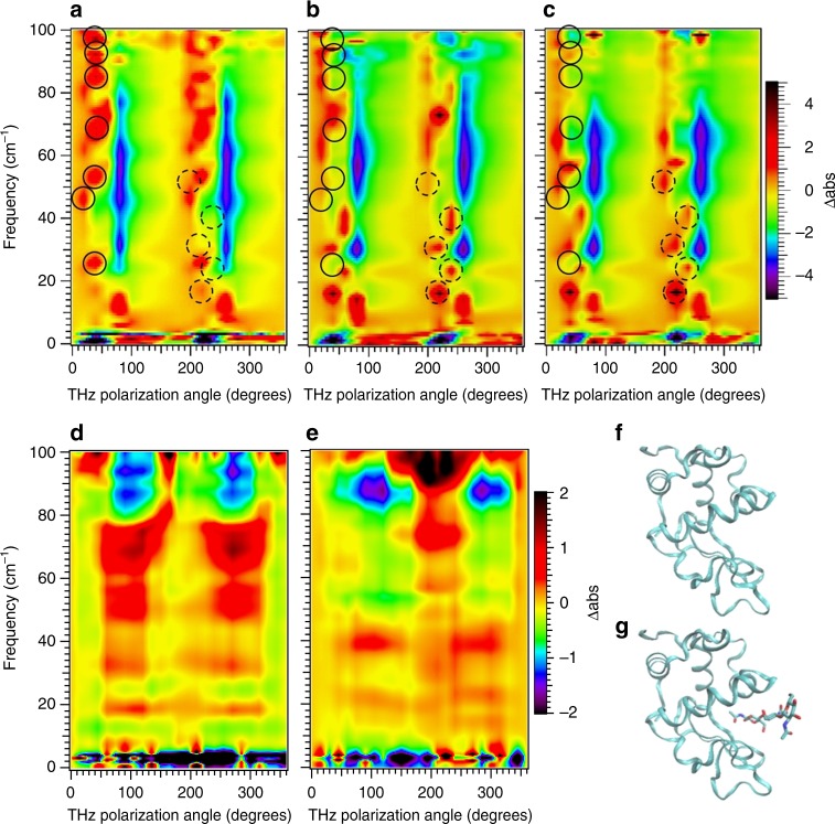
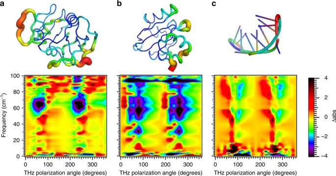
Protein structural vibrations impact biology by steering the structure to functional intermediate states; enhancing tunneling events; and optimizing energy transfer. Strong water absorption and a broad continuous vibrational density of states have prevented optical identification of these vibrations. Recently spectroscopic signatures that change with functional state were measured using anisotropic terahertz microscopy. The technique however has complex sample positioning requirements and long measurement times, limiting access for the biomolecular community. Here we demonstrate that a simplified system increases spectroscopic structure to dynamically fingerprint biomacromolecules with a factor of 6 reduction in data acquisition time. Using this technique, polarization varying anisotropy terahertz microscopy, we show sensitivity to inhibitor binding and unique vibrational spectra for several proteins and an RNA G-quadruplex. The technique's sensitivity to anisotropic absorbance and birefringence provides rapid assessment of macromolecular dynamics that impact biology.


Conflict of interest statement

The authors declare no competing interests.

Figures

Fig. 1
Fig. 1
Directionality of global intramolecular vibrations. The vector diagrams of two calculated low frequency vibrations for CEWL at (a) 12.3 cm−1 (0.37 THz) and b 11.6 cm−1 (0.35 THz). The orange shaded region highlights the binding region. While the two vibrations nearly overlap in energy, their motions are dramatically different. For the 12.3 cm−1 case the largest amplitude motion is largely out of the page, with little motion around the binding site, whereas the 11.3 cm−1 vibration moves in the page with a large compressive motion into the binding region. The black arrows indicate the light polarization direction needed to excite the vibration, demonstrating that polarization dependent absorption measurements on aligned samples readily discriminates between the two vibrations
Fig. 2
Fig. 2
Schematic of PV-ATM optical system. Sample rotation and X-Y imaging at each rotation is eliminated by THz polarization rotation
Fig. 3
Fig. 3
Relationship of PV-ATM data to vibrational absorbance for sucrose. The measured Δabs(ν,θ) using (a) far-field orientation dependent absorption, b near field anisotropic absorbance (ATM), and c PV-ATM. The THz radiation polarized in the a-b plane and the 0° orientation corresponds to the THz polarization parallel to the a axis. Also shown is simulation of the PV-ATM measurement (d). The simulations indicate that the increased structure in the PV-ATM spectra compared to anisotropic absorption arises from sensitivity to both the resonant anisotropic birefringence and the EO detector crystal polarization dependence
Fig. 4
Fig. 4
PV-ATM sensitivity to inhibitor binding. a CEWL; b CEWL-3NAG crystal 1, and c CEWL-3NAG crystal 2. The spectra is reproducible crystal to crystal. ATM measurements are shown for comparison (d) CEWL and e CEWL-3NAG. The simpler and faster PV-ATM technique provides spectra with increased structure over the ATM method. For the PV-ATM results solid circles indicate spectral features that are present in free CEWL and absent in inhibitor bound CEWL. Dashed circles indicate features present for inhibitor bound CEWL and absent for free CEWL. These are offset by 180° from the solid circles since all features have 180° periodicity from the regular alignment of the molecules. The large changes with inhibitor binding indicate the features arise from intramolecular vibrations and not crystal phonons. The crystal phonons are dependent on the intermolecular surface contacts and molecular mass, which change only slightly with binding as indicated by the X-ray structures for f free (1bwh.pdb) and for g inhibitor bound (1hew.pdb)
Fig. 5
Fig. 5
Unique dynamical fingerprints for different biomacromolecules. PV-ATM spectra for a E. coli DHFR (1dra.pdb); b photoactive yellow protein (2phy.pdb) and c RNA G-quadruplex (4xk0.pdb) rendered with Pymol B-factor putty. B-factor varies from high (red) to low (blue). While the vibrational density of states, that is the vibrational energy spectrum, for the three systems are highly similar, the actual motion for a given vibrational energy is structure specific leading to large differences in the anisotropic response

Comment in

References

    1. Austin RH, Beeson KW, Eisenstein L, Frauenfelder H, Gunsalus IC. Dynamics of Ligand Binding to Myoglobin. Biochemistry. 1975;14:5355–5373. doi: 10.1021/bi00695a021. - DOI - PubMed
    1. Bhabha G, et al. Divergent evolution of protein conformational dynamics in dihydrofolate reductase. Nat. Struct. Mol. Biol. 2013;20:1243–U1262. doi: 10.1038/nsmb.2676. - DOI - PMC - PubMed
    1. Toprak E, et al. Evolutionary paths to antibiotic resistance under dynamically sustained drug selection. Nat. Genet. 2012;44:101–U140. doi: 10.1038/ng.1034. - DOI - PMC - PubMed
    1. Acbas, G., Niessen, K. A., Snell, E. H. & Markelz, A. G. Optical measurements of long-range protein vibrations. Nat. Commun. 5, 3076 (2014). - PubMed
    1. Turton, D. A. et al. Terahertz underdamped vibrational motion governs protein-ligand binding in solution. Nat Commun. 5, 3999 (2014). - PubMed

Publication types