. 2019 Mar 4;9(1):3357.
doi: 10.1038/s41598-019-39769-z.

Deletion of Limk1 and Limk2 in mice does not alter cochlear development or auditory function

Affiliations

Deletion of Limk1 and Limk2 in mice does not alter cochlear development or auditory function

Qiaojun Fang et al. Sci Rep. .

Abstract

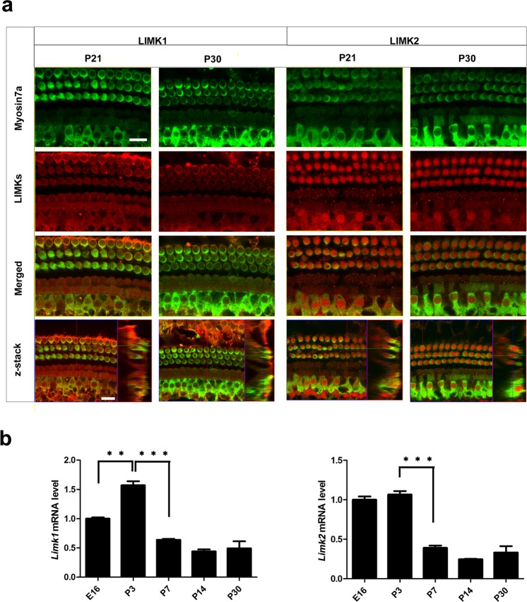
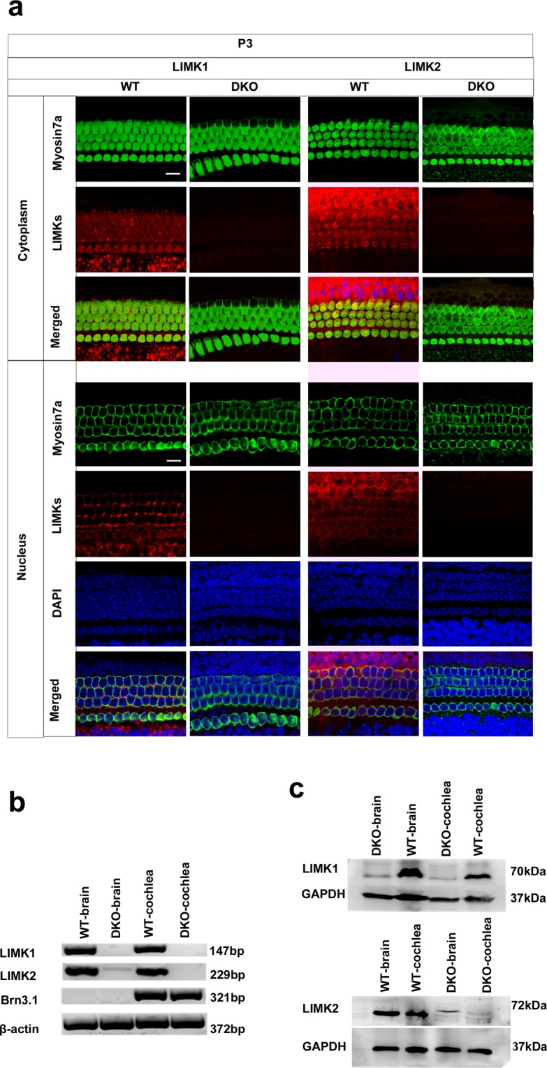
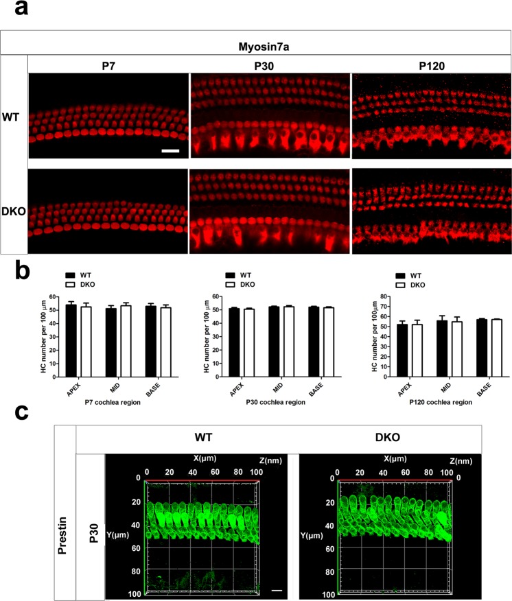
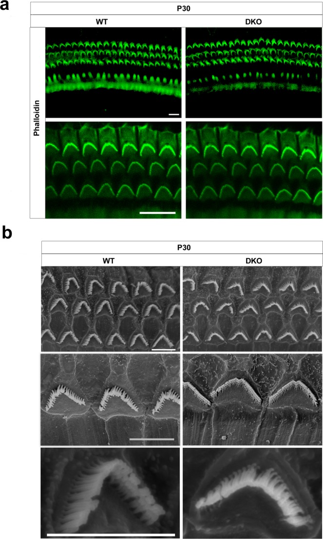
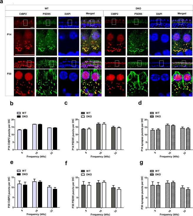
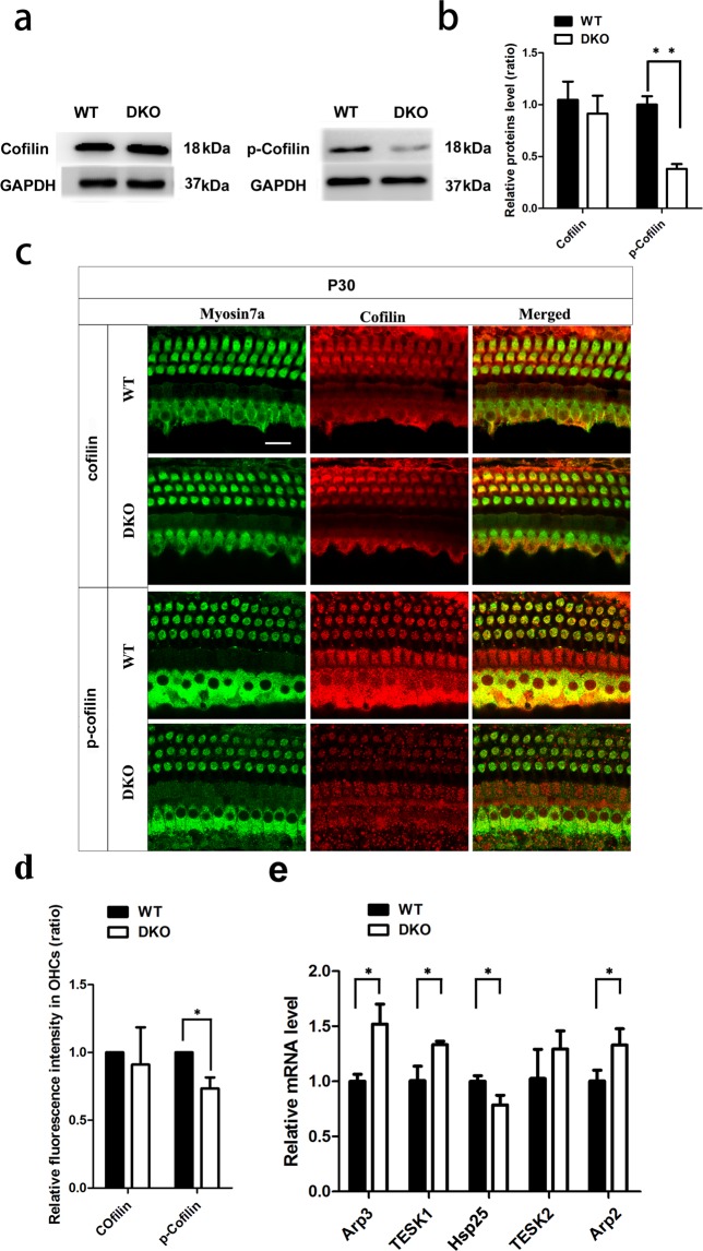
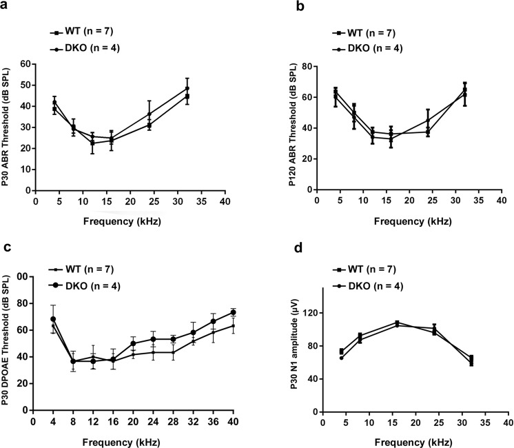
Inherited hearing loss is associated with gene mutations that result in sensory hair cell (HC) malfunction. HC structure is defined by the cytoskeleton, which is mainly composed of actin filaments and actin-binding partners. LIM motif-containing protein kinases (LIMKs) are the primary regulators of actin dynamics and consist of two members: LIMK1 and LIMK2. Actin arrangement is directly involved in the regulation of cytoskeletal structure and the maturation of synapses in the central nervous system, and LIMKs are involved in structural plasticity by controlling the activation of the actin depolymerization protein cofilin in the olfactory system and in the hippocampus. However, the expression pattern and the role of LIMKs in mouse cochlear development and synapse function also need to be further studied. We show here that the Limk genes are expressed in the mouse cochlea. We examined the morphology and the afferent synapse densities of HCs and measured the auditory function in Limk1 and Limk2 double knockout (DKO) mice. We found that the loss of Limk1 and Limk2 did not appear to affect the overall development of the cochlea, including the number of HCs and the structure of hair bundles. There were no significant differences in auditory thresholds between DKO mice and wild-type littermates. However, the expression of p-cofilin in the DKO mice was significantly decreased. Additionally, no significant differences were found in the number or distribution of ribbon synapses between the DKO and wild-type mice. In summary, our data suggest that the Limk genes play a different role in the development of the cochlea compared to their role in the central nervous system.


Conflict of interest statement

The authors declare no competing interests.

Figures

Figure 1
Figure 1
Expression of LIMKs in WT mouse cochlea. (a) Immunofluorescence staining showed that LIMK1 and LIMK2 were expressed in the cochlear epithelium in the P21 and P30 mice. Myosin7a was used as a marker for HCs. Images were taken from the basal turn of the sensory epithelium. There was no difference in the immunolabeling of LIMK1 and LIMK2 from the apical to basal turns. Scale bar = 10 µm. (b) RT-qPCR results show the changes in expression of Limks in the mouse cochlea from embryonic development to adult. β-actin was used as the internal control. Data are presented as mean ± SD. (p < 0.01, n = 5).
Figure 2
Figure 2
Analysis of LIMK expression in the DKO mice at the mRNA and protein level. (a) The cochleae of P3 DKO mice were immunolabeled with LIMK1 and LIMK2 antibodies. Scale bar = 10 µm. Images were taken from the basal turn of the sensory epithelium. There was no difference in the immunolabeling of LIMK1 and LIMK2 from the apical to basal turns. (b) RT-PCR was performed to analyze the DKO mice. Total cochlear RNA was extracted from P3 DKO and WT mice. Brn3.1 was used as the positive control, and β-actin was used as the internal control. (c) Western blot was performed to analyze DKO mice using antibodies against LIMK1 and LIMK2. Proteins from the brain and cochlea were extracted from P3 DKO and WT mice, and GAPDH was used as the internal control.
Figure 3
Figure 3
The auditory HCs are morphologically normal in the DKO mice. (a) Auditory HCs of P7, P30, and P120 mice were stained with antibodies against myosin7a and imaged using a confocal microscope. Scale bar = 10 µm. Images were taken from the basal turn of the cochlea. There was no difference in the staining from the apical to basal turns. (b) The HCs were counted and compared with age-matched WT mice (p > 0.05, n = 4). Data are presented as mean ± SD. (c) Auditory OHCs of P30 mice were stained with antibodies against prestin and imaged using a confocal microscope. Images were taken from the basal turn of the cochlea. Scale bar = 10 µm.
Figure 4
Figure 4
The auditory HC stereocilia are morphologically normal in DKO mice. (a) Auditory HC stereocilia of DKO and WT mice were stained with FITC-conjugated phalloidin and imaged using a confocal microscope. Images were taken from the basal turn of the cochlea, and there was no difference from the apical to basal turns. Scale bar = 10 µm. (b) Low magnification and high magnification scanning electron microscope images of OHC stereocilia bundles of DKO and WT mice. Images were taken from the middle turn of P30 mice. Scale bar = 5 µm.
Figure 5
Figure 5
The ribbon synapses were normal in DKO mice. (a) Ribbon synapses of P14 and P30 DKO and WT mice were stained with the ribbon synapse-specific markers CtBP2 and PSD95 and imaged under a confocal microscope. Images were taken from the basal turn of the cochlea, and there was no difference from the apical to basal turns (b,c,e,f). The total numbers of synapses from the 8-kHz to 32-kHz region were counted and compared between WT and DKO mice (d,g). The numbers of functional synapses in DKO mice were compared with WT mice. No significant differences were seen for any measurements (p > 0.05, n = 4). Scale bar = 10 µm. Data are presented as mean ± SD.
Figure 6
Figure 6
The effect of loss of LIMKs on cofilin. (a) Western blot was performed to detect the expression level of cofilin and p-cofilin in both P30 WT and DKO mice, n = 3. (b) Quantification of the Western blot (p < 0.01). (c) Auditory HCs of P30 DKO and WT mice were stained with cofilin and p-cofilin antibodies. Images were taken from the basal turn of the cochlea, and there was no difference from the apical to basal turns. Scale bar = 10 µm. (d) Quantification of cofilin and p-cofilin immunolabeling in the basal turns of OHCs (p < 0.05) n = 3. (e) RT-qPCR was performed with P30 WT and DKO mice cochleae (p < 0.05, n = 5). Data are presented as mean ± SD.
Figure 7
Figure 7
Auditory measurements show normal auditory function in DKO mice. (a) ABR thresholds of P30 DKO and WT mice were measured at 4, 8, 12, 16, 24, and 32 kHz. (b) ABR thresholds of P120 DKO and WT mice were measured at 4, 8, 12, 16, 24, and 32 kHz. (c) DPOAE thresholds were measured in response to tone bursts of 4, 8, 16, 20, 24, 28, 32, 36, and 40 kHz in P30 DKO and WT mice. (d) CAP amplitudes of P30 DKO and WT mice were measured in response to tone bursts of 4, 8, 16, and 32 kHz at 90 dB SPL. No significant differences were observed between DKO and WT mice (p > 0.05). Data are presented as mean ± SD.

References

    1. Fekete DM, Muthukumar S, Karagogeos D. Hair cells and supporting cells share a common progenitor in the avian inner ear. The Journal of neuroscience: the official journal of the Society for Neuroscience. 1998;18:7811–7821. doi: 10.1523/JNEUROSCI.18-19-07811.1998. - DOI - PMC - PubMed
    1. Li A, et al. Novel compounds protect auditory hair cells against gentamycin-induced apoptosis by maintaining the expression level of H3K4me2. Drug Delivery. 2018;25:1033–1043. doi: 10.1080/10717544.2018.1461277. - DOI - PMC - PubMed
    1. Liu W, et al. Wnt Signaling Activates TP53-Induced Glycolysis and Apoptosis Regulator and Protects Against Cisplatin-Induced Spiral Ganglion Neuron Damage in the Mouse Cochlea. Antioxid Redox Signal. 2018 doi: 10.1089/ars.2017.7288. - DOI - PubMed
    1. Savary E, et al. Cochlear stem/progenitor cells from a postnatal cochlea respond to Jagged1 and demonstrate that notch signaling promotes sphere formation and sensory potential. Mechanisms of development. 2008;125:674–686. doi: 10.1016/j.mod.2008.05.001. - DOI - PubMed
    1. Li H, et al. Meclofenamic Acid Reduces Reactive Oxygen Species Accumulation and Apoptosis, Inhibits ExcessiveAutophagy, and Protects Hair Cell-Like HEI-OC1 Cells From Cisplatin-Induced Damage. Frontiers in cellular neuroscience. 2018;12:139. doi: 10.3389/fncel.2018.00139. - DOI - PMC - PubMed

Publication types

LinkOut - more resources