. 2019 Mar 4;9(1):3361.
doi: 10.1038/s41598-019-40109-4.

Insights into individual variations in nematocyst venoms from the giant jellyfish Nemopilema nomurai in the Yellow Sea

Affiliations

Insights into individual variations in nematocyst venoms from the giant jellyfish Nemopilema nomurai in the Yellow Sea

Yang Yue et al. Sci Rep. .

Abstract

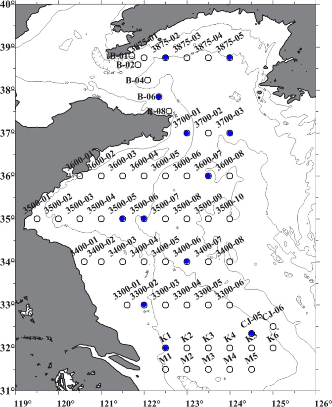
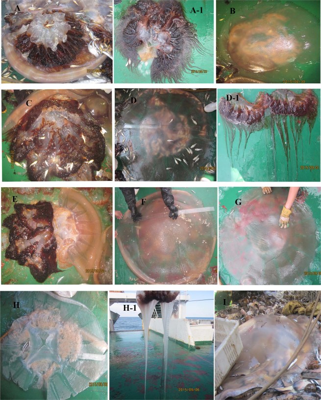
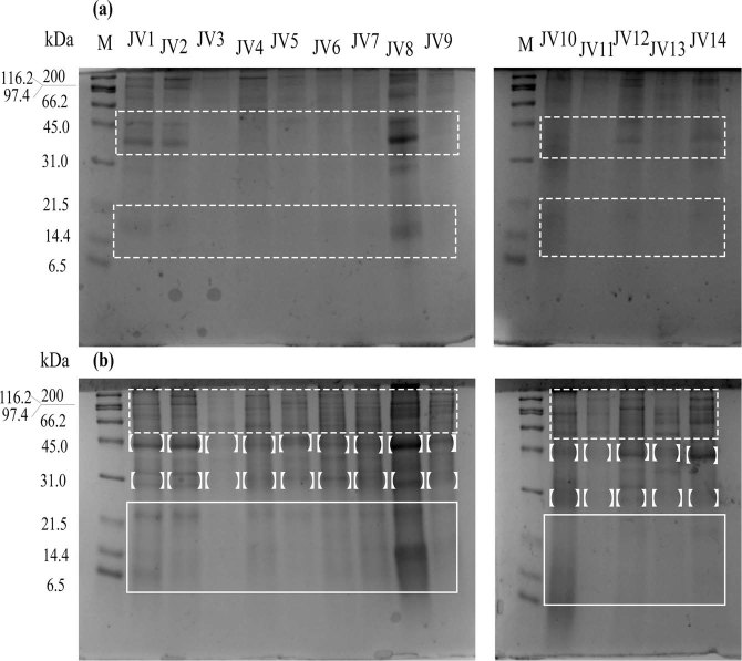
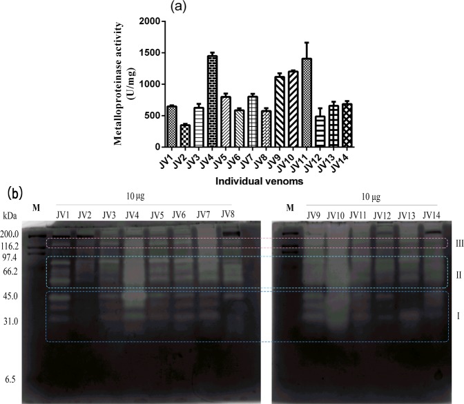
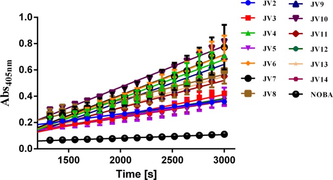
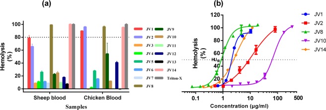
The giant jellyfish, Nemopilema nomurai, is widely distributed from the Eastern China Sea to the northern part of the Yellow Sea and has resulted in numerous hospitalizations in coastal areas of China, especially in Northern China. Our previous studies have revealed sting-related proteins in the venom of the jellyfish N. nomurai by using experimental and omics-based approaches; however, the variable symptoms of patients who have been stung by N. nomurai are not fully understood. This limited knowledge led to an examination of whether intraspecific variations occur in the venom of different N. nomurai. In the present study, 13 specimens of N. nomurai were collected from the Yellow Sea, and their venom was characterized by profiling differences in biochemical properties and biological activities. SDS-PAGE analysis presented recognizable differences in the number, intensity and presence of some protein bands. Moreover, enzymatic assays revealed considerable quantitative variations in metalloproteinase activity and PLA2-like activity. In particular, zymography assays of proteases demonstrated the general presence of abundant metalloproteinases in jellyfish nematocyst venom; however, the catalytic activities varied greatly among some specific metalloproteinases in the 28-46 kDa or 57-83 kDa range. Hemolytic assays using sheep erythrocytes suggested a predominant variance in the toxicities of different individual jellyfish venoms, with the difference between the most hemolytic and the least hemolytic venom as large as 77-fold. The current data suggested remarkable variations in the nematocyst venoms of individual N. nomurai jellyfish. These observations will provide a new understanding of the clinical manifestations induced by N. nomurai jellyfish stings and will therefore have important implications for preventing and treating jellyfish envenomations.


Conflict of interest statement

The authors declare no competing interests.

Figures

Figure 1
Figure 1
Collection stations in the Yellow Sea used by research vessel Beidou in the summer of 2015. Black circles with blue dots represent the 12 working stations from which 13 individual jellyfish were collected.
Figure 2
Figure 2
Representative photographs of individual Nemopilema nomurai jellyfish (AI) and their perspective tentacle tissues (A-1, D-1, H-1). (A) Collected at station K1; (B), station CJ-05; (C), station 3300-02; (D), station 3400-06; (E), station 3500-06; (F), station 3600-07; (G), station 3700-03; (H), station B-06; (I), station 3875-02. All photographs were taken by the author Yang Yue.
Figure 3
Figure 3
SDS-PAGE profiles of the nematocyst venoms of Nemopilema nomurai jellyfish. A total of 10 μg of venom proteins from individual jellyfish J1-J14 were loaded onto the gels and electrophoresis was performed at 120 V under nonreducing conditions (a) or reducing conditions (b) JV1-JV14 represent nematocyst venoms from jellyfish individuals J1-J14; M, protein markers (kDa). The differential bands were indicated by square brackets and rectangles.
Figure 4
Figure 4
Metalloproteinase activity. (a) Specific activity of JV1-JV14 determined using azocasein with results expressed as U/mg from at least three replicates. (b) Protease zymogram of JV1-JV14. Approximately 10 μg of each venom was loaded onto the substrate gel. The presence of clear translucent bands against the blue background indicates the existence of proteolytic enzymes. The zymogram is displayed in grayscale. I, 28–46 kDa; II, 57–83 kDa; III, 139 kDa; M, marker (kDa).
Figure 5
Figure 5
PLA2-like activity of different jellyfish nematocyst venoms assayed with NOBA. A total of 25 μL of JV1-JV14 was added to 200 μL of assay buffer containing 50 mM Tris-HCl, 5 mM CaCl2, and 100 mM NaCl, at pH 8.0 in a 96-well plate. The reactions were initiated by adding 25 μL of 1 mg/mL NOBA solution, and the absorbance was recorded for 50 min at 405 nm at 37 °C.
Figure 6
Figure 6
Hemolytic activity. (a) The hemolytic activity of JV1-JV14 against two types of blood cells, sheep erythrocytes and chicken erythrocytes at 7.0–37.6 μg/mL. A total of 50 μL of venom proteins (35.0–188.0 μg/mL in PBS) from jellyfish individuals J1-J13 was added to 200 μL of PBS-diluted erythrocytes in triplicate and then incubated at 37 °C for 30 min. The hemolytic activity was measured by examining the absorbance at 540 nm. A solution of 1% Triton X-100 in PBS and PBS alone represented 100% and 0% lysis, respectively. The hemolysis rate (%) was calculated as the percentage relative to complete lysis. (b) Concentration-response curves of JV1, JV2, JV8, JV10, and JV14 against 1% sheep erythrocytes. The results were expressed as the mean ± S.E.M. (n = 3). The dashed line indicates the HU50 values, which was defined as the concentration of protein that causes 50% lysis of sheep erythrocytes.
Figure 7
Figure 7
Cluster results of protein concentration and metalloproteinase and PLA2 activities of venom from 13 individual N. nomurai using a hierarchical clustering method. Blue, green and red outlines indicate the three groups that resulted from clustering analysis.

References

    1. Dong Z, Liu D, Keesing JK. Jellyfish blooms in China: Dominant species, causes and consequences. Marine pollution bulletin. 2010;60:954–963. doi: 10.1016/j.marpolbul.2010.04.022. - DOI - PubMed
    1. Sun S, Sun X-x, Jenkinson IR. Preface: Giant jellyfish blooms in Chinese waters. Hydrobiologia. 2015;754:1–11. doi: 10.1007/s10750-015-2320-3. - DOI
    1. Cegolon L, Heymann WC, Lange JH, Mastrangelo G. Jellyfish stings and their management: A review. Marine Drugs. 2013;11:523–550. doi: 10.3390/md11020523. - DOI - PMC - PubMed
    1. Uri S, Marina G, Liubov G. Severe delayed cutaneous reaction due to Mediterranean jellyfish (Rhopilema nomadica) envenomation. Contact Dermatitis. 2005;52:282–283. doi: 10.1111/j.0105-1873.2005.00582.x. - DOI - PubMed
    1. Qin, S., Zhang, M. & Li, M. Nematocyst dermatitis of jellyfish Stomolophus nomurai. Acta Aacademiae Medicinae Qingdao Universitatis, 1–4 (1987).

Publication types

LinkOut - more resources