. 2019 Mar 4;9(1):3351.
doi: 10.1038/s41598-019-40238-w.

p300/CBP-associated factor promotes autophagic degradation of δ-catenin through acetylation and decreases prostate cancer tumorigenicity

Affiliations

p300/CBP-associated factor promotes autophagic degradation of δ-catenin through acetylation and decreases prostate cancer tumorigenicity

Rui Zhou et al. Sci Rep. .

Abstract

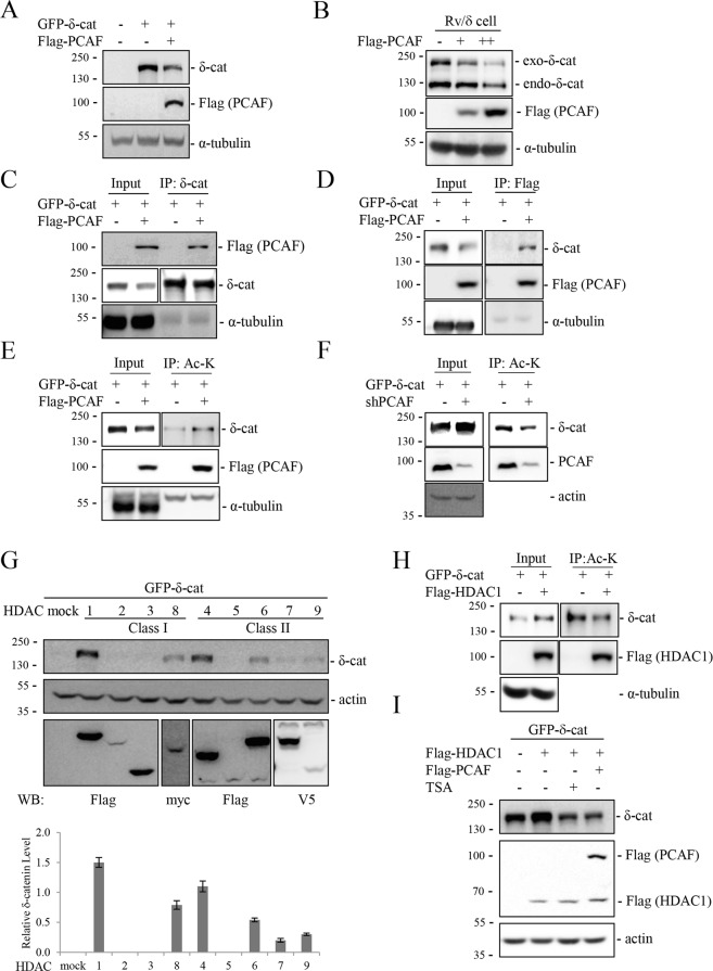
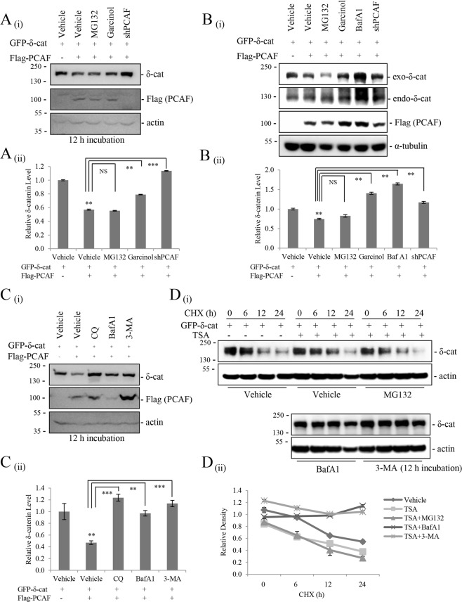
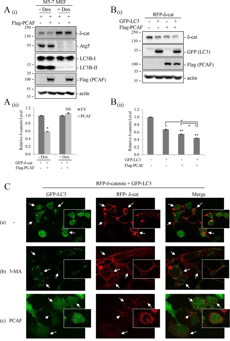
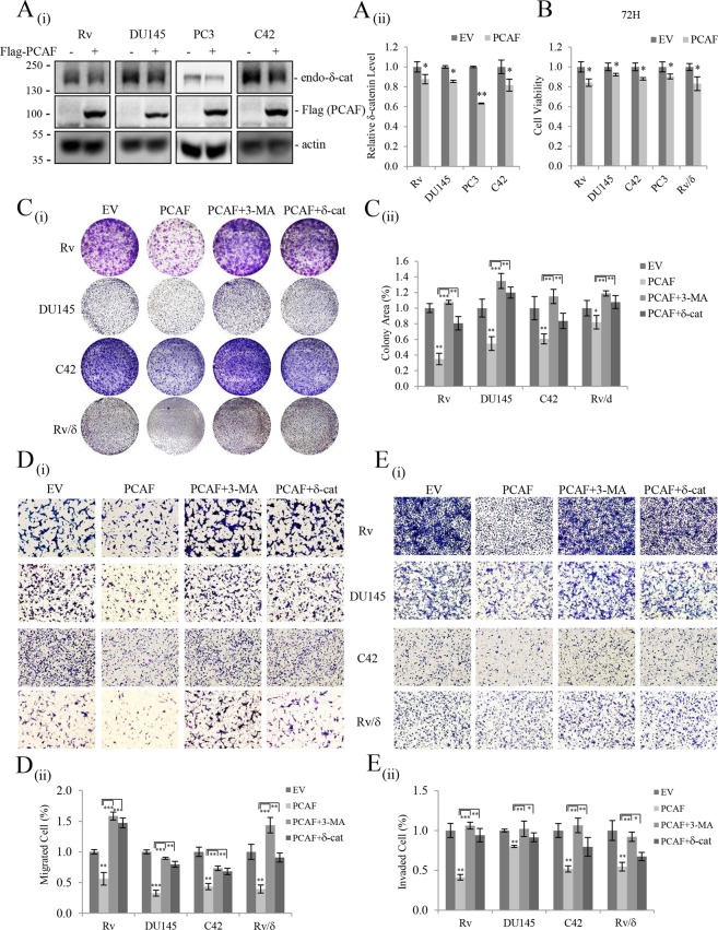
δ-Catenin shares common binding partners with β-catenin. As acetylation and deacetylation regulate β-catenin stability, we searched for histone acetyltransferases (HATs) or histone deacetylases (HDACs) affecting δ-catenin acetylation status and protein levels. We showed that p300/CBP-associated factor (PCAF) directly bound to and acetylated δ-catenin, whereas several class I and class II HDACs reversed this effect. Unlike β-catenin, δ-catenin was downregulated by PCAF-mediated acetylation and upregulated by HDAC-mediated deacetylation. The HDAC inhibitor trichostatin A attenuated HDAC1-mediated δ-catenin upregulation, whereas HAT or autophagy inhibitors, but not proteasome inhibitors, abolished PCAF-mediated δ-catenin downregulation. The results suggested that PCAF-mediated δ-catenin acetylation promotes its autophagic degradation in an Atg5/LC3-dependent manner. Deletions or point mutations identified several lysine residues in different δ-catenin domains involved in PCAF-mediated δ-catenin downregulation. PCAF overexpression in prostate cancer cells markedly reduced δ-catenin levels and suppressed cell growth and motility. PCAF-mediated δ-catenin downregulation inhibited E-cadherin processing and decreased the nuclear distribution of β-catenin, resulting in the suppression of β-catenin/LEF-1-mediated downstream effectors. These data demonstrate that PCAF downregulates δ-catenin by promoting its autophagic degradation and suppresses δ-catenin-mediated oncogenic signals.


Conflict of interest statement

The authors declare no competing interests.

Figures

Figure 1
Figure 1
PCAF and HDACs regulate the acetylation status and levels of δ-catenin. (A,B) PCAF decreases δ-catenin levels. HEK293T (A) and δ-catenin-overexpressing CWR22Rv-1 (Rv/δ) (B) cells were transfected as indicated, and cell lysates were subjected to immunoblotting. (CF) PCAF interacts with and acetylates δ-catenin. HEK293T cells were transfected as indicated, and cell lysates were subjected to immunoprecipitation with anti-δ-catenin (C), anti-Flag (D), or anti-acetylated-lysine (E,F), followed by immunoblotting of precipitated proteins. (GI) HDACs deacetylate and upregulate δ-catenin. HEK293T cells were transfected as indicated, and cell lysates were subjected to immunoblotting (G) or immunoprecipitation with anti-acetylated-lysine (H). At 12 h post-transfection of HEK293T cells with GFP-δ-catenin and HDAC1, cells were treated with 0.2 µM trichostatin A (TSA) or transfected with Flag-PCAF, and incubated for 24 h, followed by immunoblotting with anti-δ-catenin and anti-Flag antibodies (I). α-Tubulin or ß-actin was used as a loading control. Relative δ-catenin/actin/HDACs ratios from three different experimental results are shown as a bar graph (G). Values are presented as the mean ± SEM. “−”, Mock transfection or vehicle treatment.
Figure 2
Figure 2
PCAF-mediated δ-catenin acetylation promotes autophagic degradation of δ-catenin. (A,B) The acetyltransferase activity of PCAF is required for the downregulation of δ-catenin. However, proteasome inhibition does not suppress the effect of PCAF on downregulating δ -catenin. HEK293Tcells transfected with GFP-δ-catenin and Flag-PCAF (A) and Rv/δ cells transfected with Flag-PCAF (B) were treated with the proteasome inhibitor MG132 (10 μM) or the histone acetyltransferase inhibitor Garcinol (5 µM) or Baf A1 (100 nM) or transfected with shPCAF and incubated for 12 h, and then cell lysates were subjected to immunoblotting. (C) Autophagy inhibitors attenuate PCAF-mediated δ-catenin degradation. HEK293T cells were transfected with the indicated plasmids. At 12 h post-transfection, cells were treated with the autophagy inhibitors chloroquine (CQ, 100 μM), bafilomycin A1 (BafA1, 100 nM), or 3-methyladenine (3-MA, 5 mM), and cell lysates were subjected to immunoblotting. (D) Autophagy inhibitors increase the stability of δ-catenin. HEK293T cells transfected with δ-catenin and treated with 0.2 µM TSA were treated with MG132 (10 μM) or Baf A1 (100 nM) or 3-MA (5 mM) and incubated for 12 h, and then treated with cycloheximide (CHX, 20 ng/ml) for indicated time (h), and then cell lysates were subjected to immunoblotting. α-Tubulin or ß-actin was used as a loading control. Relative δ-catenin/actin ratios from at least three independent experiments are shown as a bar graph in each panel (ii). Values are presented as the mean ± SEM. **p < 0.01; ***p < 0.001; NS, no significant difference compared with the control group. “−”, Mock transfection or vehicle treatment.
Figure 3
Figure 3
The Atg5/12-LC3 pathway is indispensable for PCAF-mediated δ-catenin degradation. (A) Atg5 is necessary for PCAF-mediated δ-catenin degradation. Tet-off Atg5−/− mouse embryonic fibroblasts m5-7 were cultured in the presence or absence of 10 ng/ml doxycycline (Dox) for 4 days. Then, cells were transfected as indicated for 24 h, and cell lysates were subjected to immunoblotting. (B) Overexpression of LC3 further decreases δ-catenin levels. HEK293T cells were transfected as indicated, and cell lysates were subjected to immunoblotting. Actin was used as a loading control. Relative values of δ-catenin/actin ratios from at least three independent experiments are shown as a bar graph in each panel (ii). Values are presented as the mean ± SEM. *p < 0.05; **p < 0.01; NS, no significant difference compared with the control group. (C) The cellular localization of δ-catenin depends on autophagic activity and PCAF. HEK293T cells were transiently transfected with a plasmid encoding GFP-LC3 and RFP-δ-catenin for 12 h, and incubated in the absence (a) or presence of 3-MA (b) or transfected with flag-PCAF (c) for 24 h. (a) Arrows indicate δ-catenin located at the plasma membrane or cell-cell contact region. (b) Arrows indicate cytoplasmic δ-catenin accumulation. 3-MA treatment dispersed LC3 signals and no colocalization with δ-catenin was observed. (c) Arrows indicated δ-catenin colocalization with LC3. PCAF overexpression decreased the accumulation of plasma membrane or cell-cell contact region and increased the colocalization with LC3. “−”, Mock transfection and/or vehicle treatment.
Figure 4
Figure 4
Multiple domains of δ-catenin are responsible for PCAF-mediated δ-catenin downregulation. (A) Schematic diagram of different constructs of full length GFP-δ-catenin and its deletion mutants (ΔC207, ΔN85–325, ΔN&ΔC, 1–690, 691–1040, 1–640, 1–600, 1–579, and 1–499) and the distribution of Lys residues on these deletion mutants. Lys residues (Lys1049, Lys1050, and Lys1158) form the ubiquitin-dependent degron of δ-catenin. (B) Both 1–690 and 691–1040 deletion mutants of δ-catenin were downregulated by PCAF. HEK293T cells were transfected with different combinations of the indicated plasmids, and cell lysates were subjected to immunoblotting. Actin was used as a loading control. Relative values of δ-catenin/actin ratios from at least three independent experiments are shown as a bar graph in each panel (ii). Values are presented as the mean ± SEM. *p < 0.05; **p < 0.01; ***p < 0.001; NS, no significant difference compared with the control group. “−”, Mock transfection.
Figure 5
Figure 5
Multiple lysine residues in the N-terminus are responsible for PCAF-mediated δ-catenin downregulation. (A) Schematic representation of the triple arginine mutation at Lys360, Lys371, and Lys428 (FL KR), and the deletion/arginine mutation constructs of δ-catenin 1–499, 1–499 KR, 85–499, 85–499 KR, 1–499∆N KR, and 325–499 KR. (B) Deletion mutants 85–499 KR and 325–499 KR of δ-catenin were not affected by PCAF. HEK293T cells were transfected with the indicated plasmids expressing δ-catenin constructs, and cell lysates were subjected to immunoblotting with anti-GFP and anti-Flag antibody. (C) 3-MA restored the downregulated FL KR, 85–499, and 1–499∆N KR mutants of δ-catenin except the 85–499 KR mutation. HEK293T cells were transfected with the indicated plasmids expressing δ-catenin constructs and incubated with 3-MA (1 mM) for 24 h, and cell lysates were subjected to immunoblotting with anti-GFP and anti-Flag antibodies. (D) PCAF did not acetylate δ-catenin 85–499 KR mutation. HEK293T cells were transfected with full length GFP-δ-catenin or 85–499 KR mutant together with or without Flag-PCAF, and each cell lysates were subjected to immunoprecipitation with anti-acetylated-lysine, followed by immunoblotting of precipitated proteins. α-Tubulin or ß-actin was used as a loading control. Relative values of δ-catenin/actin ratios from at least three independent experiments are shown as a bar graph in each panel (ii). Values are presented as the mean ± SEM. *p < 0.05; **p < 0.01; NS, no significant difference compared with the control group. “−”, Mock transfection or vehicle treatment.
Figure 6
Figure 6
PCAF-mediated δ-catenin downregulation suppresses prostate cancer cell growth and motility. (A) PCAF reduced endogenous δ-catenin levels in Rv, DU145, PC3, and C42 prostate cancer cell lines. Four prostate cancer cell lines were transfected with empty vector or Flag-PCAF and subjected to immunoblotting with anti-δ-catenin or anti-Flag antibody. (BC) PCAF suppressed the viability and growth of Rv, DU145, PC3, C42, and Rv/δ cells, and 3-MA treatment or δ-catenin overexpression restored the decreased growth of PCAF-overexpressing prostate cancer cells. Cell viability (B) and cell growth (C) were determined by the MTT assay and clonogenic assay, respectively, in empty vector or Flag-PCAF transfected cells treated or transfected with vehicle or 3-MA or δ-catenin. (DE) PCAF reduced the migration and invasion abilities of Rv, DU145, C42, and Rv/δ cells while 3-MA treatment or δ-catenin overexpression restored the decreased migration and invasion of PCAF-overexpressing prostate cancer cells. Cells were transfected with Flag-PCAF together with or withiout δ-catenin or empty vector, and migration and invasion assays were performed in the presence of vehicle or 3-MA with Transwell chambers coated without (D) or with (E) gelatin, respectively. Quantitative analysis of δ-catenin levels, cell viability, colony area, and migrated and invaded cell numbers from at least three independent experiments are shown as a bar graph in each panel (ii). Values are presented as the mean ± SEM. *p < 0.05; **p < 0.01; ***p < 0.001; NS, no significant difference compared with the control group. “−”, Mock transfection.
Figure 7
Figure 7
PCAF inhibits E-cadherin processing and inactivates β-catenin-mediated signaling. (A) PCAF-mediated δ-catenin downregulation reduced E-cadherin processing. PCAF decreased E-cadherin fragment levels in a dose-dependent manner, whereas total E-cadherin protein levels increased. E-cadherin processing was measured in Rv/δ cells transfected with increasing doses of Flag-PCAF. (B) PCAF suppressed subcellular β-catenin distribution and its downstream effectors, c-myc and cyclin-D1. Nuclear and cytoplasmic fractionation of β-catenin, c-myc, and cyclin-D1 was performed in Rv/δ and PC3 cells transfected with Flag-PCAF. Moderate changes in total β-catenin, c-myc, and cyclin-D1 levels were also observed. α-Tubulin was used as a cytoplasmic marker, and α-histone H3 was used as a nuclear marker. Relative values from at least three independent experiments are shown as a bar graph in each panel (ii/iii). (C) PCAF decreased β-catenin target gene levels. The mRNA levels of c-myc and cyclin-D1 were analyzed by qRT-PCR in Rv/δ and PC3 cells transfected with Flag-PCAF. Values are presented as the mean ± SEM. *p < 0.05; **p < 0.01; NS, no significant difference compared with the control group. “−”, No transfection.

Similar articles

Cited by

References

    1. Shibamoto S, et al. Association ofp120, a tyrosine kinase substrate, with E-cadherin/catenin complexes. The Journal of cell biology. 1995;128:949–957. doi: 10.1083/jcb.128.5.949. - DOI - PMC - PubMed
    1. Paffenholz R, Franke WW. Identification and localization of a neurally expressed member of the plakoglobin/armadillo multigene family. Differentiation; research in biological diversity. 1997;61:293–304. doi: 10.1046/j.1432-0436.1997.6150293.x. - DOI - PubMed
    1. Lu Q, et al. Increased expression of δ-catenin/neural plakophilin-related armadillo protein is associated with the down-regulation and redistribution of E-cadherin and p120 ctn in human prostate cancer. Human pathology. 2005;36:1037–1048. doi: 10.1016/j.humpath.2005.07.012. - DOI - PubMed
    1. Shrestha H, et al. Investigation of the molecular mechanism of delta-catenin ubiquitination: Implication of beta-TrCP-1 as a potential E3 ligase. Biochimica et biophysica acta. 2016;1863:2311–2321. doi: 10.1016/j.bbamcr.2016.06.006. - DOI - PubMed
    1. Kim H, et al. delta-Catenin promotes E-cadherin processing and activates beta-catenin-mediated signaling: implications on human prostate cancer progression. Biochimica et biophysica acta. 2012;1822:509–521. doi: 10.1016/j.bbadis.2011.12.015. - DOI - PMC - PubMed

Publication types