. 2019 Feb 18:10:92.
doi: 10.3389/fphys.2019.00092. eCollection 2019.

hCLE/RTRAF-HSPC117-DDX1-FAM98B: A New Cap-Binding Complex That Activates mRNA Translation

Affiliations

hCLE/RTRAF-HSPC117-DDX1-FAM98B: A New Cap-Binding Complex That Activates mRNA Translation

Alejandra Pazo et al. Front Physiol. .

Abstract

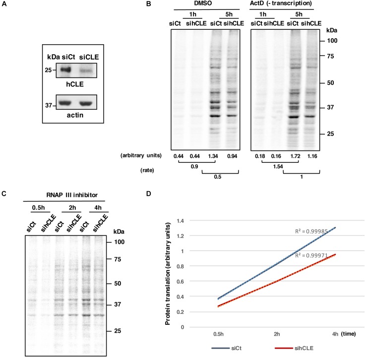
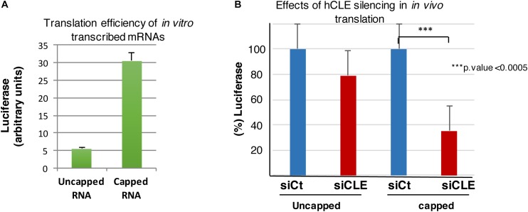
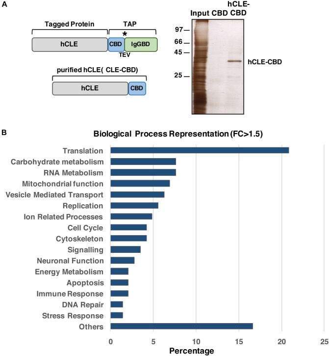
hCLE/C14orf166/RTRAF, DDX1, and HSPC117 are components of cytoplasmic mRNA-transporting granules kinesin-associated in dendrites. They have also been found in cytoplasmic ribosome-containing RNA granules that transport specific mRNAs halted for translation until specific neuronal signals renders them accessible to the translation machinery. hCLE associates to DDX1, HSPC117, and FAM98B in HEK293T cells and all four proteins bind to cap analog-containing resins. Competition and elution experiments indicate that binding of hCLE complex to cap resins is independent of eIF4E; the cap-binding factor needed for translation. Purified hCLE free of its associated proteins binds cap with low affinity suggesting that its interacting proteins modulate its cap association. hCLE silencing reduces hCLE accumulation and that of its interacting proteins and decreases mRNA translation. hCLE-associated RNAs have been isolated and sequenced; RNAs involved in mRNA translation are specifically associated. The data suggest that RNA granules may co-transport RNAs encoding proteins involved in specific functions together with RNAs that encode proteins needed for the translation of these specific RNAs and indicate an important role for hCLE modulating mRNA translation.

Keywords: cap-binding; local translation; mRNA translation; protein complexes; translation activation.


Figures

FIGURE 1
FIGURE 1
Endogenous hCLE complexes bind cap analog resins. (A) Nuclear and cytoplasmic extracts of HEK293T cells were incubated with control (4B) or cap analog (cap) resins. Western blot analysis of bound proteins using the corresponding antibodies are shown. (B) Graph showing the percentage of the indicated proteins bound to m7GTP analog resin (blue) or control resin (yellow), compared to the initial input that was incubated with the corresponding resins. Total extracts and the data of three independent experiments were used. (C) Total protein extracts from HEK293T cells were incubated with cap analog resin and eluted with 1 mM m7GTP. Eluted fractions and resin-bound proteins were resolved in non-denaturing gels and analyzed by Western blot with indicated antibodies. monomer, ∗∗dimers, and ∗∗∗trimers. Cap-binding experiments were repeated more than five times. Input:bound protein ratios were: 1:12 in total and cytoplasmic HEK293T cell extracts; 1:6 in HEK293T cell nuclear extracts.
FIGURE 2
FIGURE 2
hCLE forms oligomers. (A) Total protein extracts of HEK293T cells transfected with plasmids expressing the whole hCLE sequence (hCLEwt), N-terminal domain [hCLE(Nt)] (aa 1–120) or C-terminal domain [hCLE(Ct)] (aa 120–244) were analyzed by Western blot with anti-hCLE antibodies. (B) Silver staining of hCLE-CBD purified protein dyalized in the presence of non-reducing agents (No. Reduc.). (C) Purified hCLE was dyalized in reducing (Reduc.) or non-reducing buffer (No Reduc.) and analyzed in non-denaturing gel followed by Western blot with anti-hCLE antibodies. monomer and ∗∗dimers. 1X and 2X represent single or double amount of purified protein. Three independent experiments were done.
FIGURE 3
FIGURE 3
Biochemical properties of the hCLE-complex cap-binding activity. (A) Total extracts of HEK293T cells were incubated with cap analog (cap) or control resins (4B) in the presence of different concentrations of m7GTP, GTP or ATP (2 and 10 mM). Retained hCLE and eIF4E proteins in the corresponding resins after extensive washes were analyzed by Western blot. The experiments were repeated three times. (B) Total extracts of HEK293T cells were incubated with cap analog and control resins. After washing, protein was eluted sequentially with increasing concentrations of m7GTP, GTP or ATP (0.05, 0.1, and 1 mM). Eluted and resin-bound proteins were analyzed by Western blot. The experiments were repeated three times. (C) HEK293T cells were transfected with a control silencing plasmid (shCt) or with a plasmid specific for eIF4E silencing (sh4E). Total extracts were incubated with cap-analog resin alone or in the presence of GTP 2 μM and after washing, the retained proteins analyzed by SDS-gels and Western blots. (D) Total extracts of HEK293T cells were incubated with cap analog resins as indicated in part B. Protein was eluted sequentially with increasing concentrations of m7GTP, or m7GpppG (0.05, 0.2, and 1 mM). Eluted proteins were analyzed by Western blot and quantitated using ImageJ application (bottom). The experiments were repeated two times.
FIGURE 4
FIGURE 4
Purified hCLE binds cap analog. (A) Silver staining of Ni2+-purified His-hCLE protein. Only hCLE is visible after purification; arrows indicate molecular weight for associated proteins. (B) Western blot analysis of indicated proteins in total extracts (Input) or purified fraction of His-hCLE. To obtain the most accurate results, we analyzed all the proteins from the same sample and experiment. (C) (Top); Binding of Ni2+-purified His-hCLE proteins to cap analog and control resins. Bound protein was analyzed by Western blot. The relative amounts applied to SDS-gels were Input:resins bound 1:30. (Bottom); Purified His-hCLE was incubated with cap analog (cap) or control resins (4B) in the presence of 10 mM of GTP, or m7GTP as competitors and His-hCLE binding was analyzed by Western blot. The relative amounts applied to SDS-gels were Input:resins bound 1:7.5. The experiments were repeated at least three times. monomer.
FIGURE 5
FIGURE 5
hCLE controls RNA translation. HEK293T cells were infected with lentivirus expressing a control siRNA (siCt) or a siRNA that targets hCLE (siCLE). (A) Western blot detection of hCLE in control or silenced cells. (B) HEK293T cells were infected with control (siCt) or a siRNA that targets hCLE lentiviruses. Control or silenced HEK293T cells were cultured in Met/Cys-depleted DMEM (30 min); cells were untreated (DMSO) or actinomycin D (Act D)-treated (30 min), followed by 35S-Met-Cys addition (time 0). At the indicated times aliquots were taken and used for SDS-gels and quantitated using ImageJ application (bottom). Rate represents the differences between signals obtained at 1h after 35S-Met-Cys addition and those obtained at 5 h. A representative image is shown. The experiment was repeated three times. (C) HEK293T cells were infected with lentivirus expressing a control siRNA (siCt) or a siRNA that targets hCLE. Control or silenced HEK293T cells were cultured in Met/Cys-depleted DMEM (30 min) and then treated with 80 μM of RNAP III inhibitor followed by 35S-Met-Cys addition (time 0). At the indicated times aliquots were taken and used for SDS-gels and quantitated using ImageJ application. A representative image is shown. (D) Quantitation of data obtained from part C. The experiment was repeated two times.
FIGURE 6
FIGURE 6
hCLE controls cap-dependent translation. (A) In vitro transcribed luciferase RNA was left uncapped or capped and both RNAs used for luciferase determination by in vitro translation assay. (B) HEK293T cells were transfected with 0.5 μg/2 × 105 cells of in vitro transcribed luciferase RNA uncapped or capped and 1.5 h post-transfection luciferase activity was analyzed. The graphic shows the percentage of translated uncapped or capped luciferase RNA in control (taking as 100%) and hCLE silenced cells. Four independent experiments with three technical replicates were done. ∗∗∗p-value < 0.0005.
FIGURE 7
FIGURE 7
hCLE binds specific messenger RNAs. (A) Scheme of the hCLE-TAP fusion protein used for tandem affinity purification (TAP) and the final purified protein hCLE-CBD. Peptides bearing the calmodulin-binding domain (CBD) and the IgG-binding domain of Staphylococcus aureus protein A (IgGBD) are separated by a cysteine protease (TEV) cleavage sequence. (B) Silver staining of hCLE-CBD purified protein used for high throughout sequencing. (C) Percent representation of mRNAs associated to hCLE-CBD, grouped by biological function according to GeneOntology annotations. Two biological replicates were used. represents the TEV protease cleavage site.

Similar articles

Cited by

References

    1. Alfonso R., Lutz T., Rodriguez A., Chavez J. P., Rodriguez P., Gutierrez S., et al. (2011). CHD6 chromatin remodeler is a negative modulator of influenza virus replication that relocates to inactive chromatin upon infection. Cell. Microbiol. 13 1894–1906. 10.1111/j.1462-5822.2011.01679.x - DOI - PubMed
    1. Anders S., Pyl P. T., Huber W. (2015). HTSeq–a Python framework to work with high-throughput sequencing data. Bioinformatics 31 166–169. 10.1093/bioinformatics/btu638 - DOI - PMC - PubMed
    1. Benjamini Y., Drai D., Elmer G., Kafkafi N., Golani I. (2001). Controlling the false discovery rate in behavior genetics research. Behav. Brain Res. 125 279–284. 10.1016/S0166-4328(01)00297-2 - DOI - PubMed
    1. Burgui I., Yángüez E., Sonenber N., Nieto A. (2007). Influenza mRNA translation revisited: is the eIF4E cap-binding factor required for viral mRNA translation? J. Virol. 81 12427–12438. 10.1128/JVI.01105-07 - DOI - PMC - PubMed
    1. Castello A., Fischer B., Eichelbaum K., Horos R., Beckmann B. M., Strein C., et al. (2012). Insights into RNA biology from an atlas of mammalian mRNA-binding proteins. Cell 149 1393–1406. 10.1016/j.cell.2012.04.031 - DOI - PubMed