. 2019 May 1;316(5):E817-E828.
doi: 10.1152/ajpendo.00522.2018. Epub 2019 Mar 5.

Evidence for a role for Sestrin1 in mediating leucine-induced activation of mTORC1 in skeletal muscle

Affiliations

Evidence for a role for Sestrin1 in mediating leucine-induced activation of mTORC1 in skeletal muscle

Dandan Xu et al. Am J Physiol Endocrinol Metab. .

Abstract

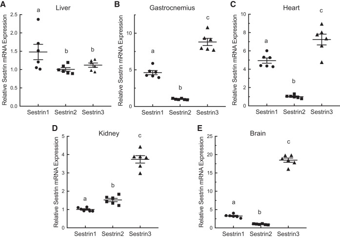
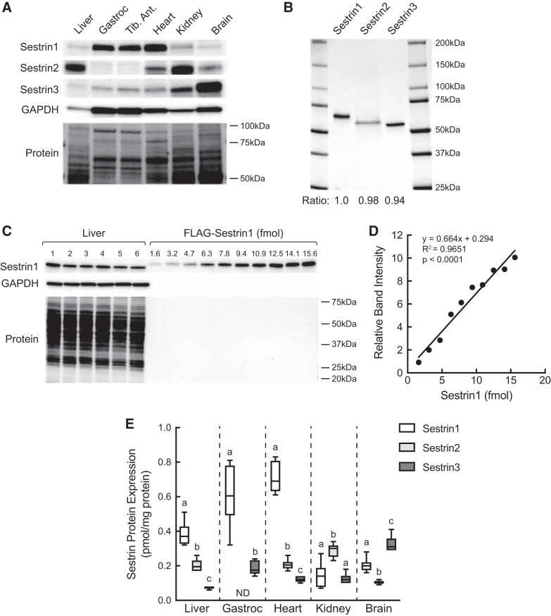
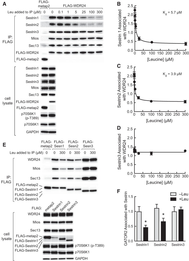
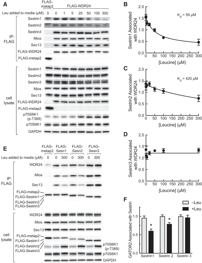
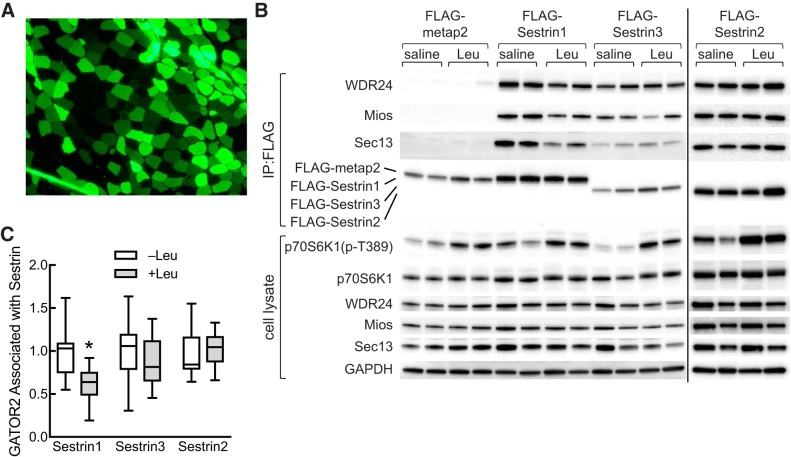
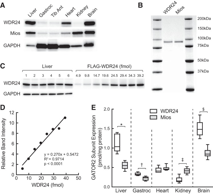
Previous studies established that leucine stimulates protein synthesis in skeletal muscle to the same extent as a complete mixture of amino acids, and the effect occurs through activation of the mechanistic target of rapamycin in complex 1 (mTORC1). Recent studies using cells in culture showed that the Sestrins bind leucine and are required for leucine-dependent activation of mTORC1. However, the role they play in mediating leucine-dependent activation of the kinase in vivo has been questioned because the dissociation constant of Sestrin2 for leucine is well below circulating and intramuscular levels of the amino acid. The goal of the present study was to compare expression of the Sestrins in skeletal muscle to other tissues and to assess their relative role in mediating activation of mTORC1 by leucine. The results show that the relative expression of the Sestrin proteins varies widely among tissues and that in skeletal muscle Sestrin1 expression is higher than Sestrin3, whereas Sestrin2 expression is markedly lower. Analysis of the dissociation constants of the Sestrins for leucine as assessed by leucine-induced dissociation of the Sestrin·GAP activity toward Rags 2 (GATOR2) complex revealed that Sestrin1 has the highest affinity for leucine and that Sestrin3 has the lowest affinity. In agreement with the dissociation constants calculated using cells in culture, oral leucine administration promotes disassembly of the Sestrin1·GATOR2 complex but not the Sestrin2 or Sestrin3·GATOR2 complex. Overall, the results presented herein are consistent with a model in which leucine-induced activation of mTORC1 in skeletal muscle in vivo occurs primarily through release of Sestrin1 from GATOR2.

Keywords: Sestrin; leucine; mTOR; skeletal muscle.


Conflict of interest statement

No conflicts of interest, financial or otherwise, are declared by the authors.

Figures

Fig. 1.
Fig. 1.
Oral administration of leucine to fasted rats differentially activates mTORC1 in liver, gastrocnemius, tibialis anterior, heart, and kidney, but not brain. Sprague-Dawley rats (200–250 g) were fasted overnight and the next morning were administered either saline or leucine (Leu) by oral gavage as described under materials and methods. Tissues were removed 45 min later, and Western blot analysis was performed as described under materials and methods for phosphorylated p70S6K1 [p70S6K1(p-T389)] and 4E-BP1 [4E-BP1(p-S65)], total p70S6K1 and 4E-BP1, and GAPDH (A). PVDF membrane was stained for protein using Pierce Reversible Stain for PVDF and then destained before Western blot analysis. The relative phosphorylation of p70S6K1 (B) and 4E-BP1 (C) is expressed as a fraction of the saline value for the respective tissue. Values represent means ± SE (n = 6). Closed bars, saline; shaded bars, leucine. *P < 0.0001 vs. respective saline condition by two-way ANOVA; ‡P < 0.0001 vs. all other leucine groups by two-way ANOVA; †P < 0.0001 vs. brain, leucine by two-way ANOVA. 4E-BP1, eukaryotic initiation factor 4E-binding protein 1; Gastroc, gastrocnemius; mTORC1, mechanistic target of rapamycin in complex 1; p70S6K1, ribosomal protein S6 protein kinase; Tib. Ant., tibialis anterior.
Fig. 2.
Fig. 2.
Sestrin1, 2, and 3 mRNA expression in liver (A), gastrocnemius (B), heart (C), kidney (D), and brain (E) of freely fed rats. RNA was extracted from tissues, and Sestrin1, 2, and 3 mRNAs were quantitated by quantitative real-time PCR as described under materials and methods. Values represent means ± SE (n = 6). Values not sharing a letter are significantly different by one-way ANOVA, P < 0.05.
Fig. 3.
Fig. 3.
Western blot analysis of Sestrin1, 2, and 3 protein expression in liver, gastrocnemius, tibialis anterior, heart, kidney, and brain. A: representative Western blot analysis of Sestrin and GAPDH expression. Membranes were stained with Pierce Reversible Stain for PVDF and then destained before Western blot analysis; a representative membrane is shown. Sestrin1, 2, and 3 antibodies were validated using HEK293T cells lacking all three Sestrin proteins [kindly provided by Dr. David Sabatini (21)]. B: FLAG-tagged Sestrin1, 2, and 3 were individually expressed in and purified from HEK293T cells as described under materials and methods. Purified proteins (2 µg) were resolved by SDS-PAGE, and the gel was stained with SimplyBlue SafeStain. Ratio, band intensity relative to Sestrin1. C: Western blot analysis of Sestrin1 and GAPDH expression in liver (n = 6) and various amounts of purified FLAG-Sestrin1. Membranes were stained for total protein loading before Western blot analysis as described above for A. D: representative standard curve for FLAG-Sestrin1. E: quantification of Western blot analysis of Sestrin1, 2, and 3 expression in various tissues. Values represent means ± SE (n = 6). Method used to quantify Sestrin1 shown in C and D was also used to quantify Sestrins 2 and 3 using the respective purified proteins (B). ND, Sestrin2 expression in gastrocnemius was too low to be accurately quantitated, and therefore is not included on the graph. For each tissue except gastrocnemius, values not sharing the same letter are significantly different by one-way ANOVA, P < 0.001. For gastrocnemius, P < 0.0005 by t-test. Gastroc, gastrocnemius; Tib. Ant., tibialis anterior.
Fig. 4.
Fig. 4.
In vitro assessment of Sestrin affinity for leucine. A: FLAG-metap2 or FLAG-WDR24 were expressed in HEK293T cells, and the next day the cells were incubated in medium lacking leucine for 5 h. FLAG-tagged proteins were immunoprecipitated from cell lysates using anti-FLAG beads as described under materials and methods. Beads were incubated for 2 h at 4°C with the concentrations of leucine indicated in the figure and then washed, and then protein bound to the beads was subjected to Western blot analysis. In addition, an aliquot of cell lysate was subjected to Western blot analysis. Representative blots are shown. BD: Western blots were subjected to densitometric analysis, and a nonlinear curve fit was performed as described under materials and methods. Values represent means ± SE (n = 3). E: FLAG-tagged metap2 or Sestrin1, 2, or 3 was expressed in HEK293T cells, and the next day the cells were incubated in medium lacking leucine for 5 h. FLAG-tagged proteins were immunoprecipitated from cell lysates using anti-FLAG beads as described under materials and methods. Beads were washed and then incubated for 2 h at 4°C with or without 300 µM leucine as indicated in the figure. In addition, an aliquot of cell lysate was subjected to Western blot analysis. Representative blots are shown. F: quantitation of Western blots from FLAG-immunoprecipitates incubated with or without 300 µM leucine. Because WDR24, Mios, and Sec13 are subunits of GATOR2, and leucine-induced changes in their association with the Sestrins were qualitatively similar, values for WDR24, Mios, and Sec13 were combined to provide an estimate for GATOR2 associated with Sestrin. Values represent the means ± SE (n = 3). *P < 0.05 vs. respective −Leu condition. GATOR2, GAP activity toward Rags 2; IP, immunoprecipitation; Kd, dissociation constant; Leu, leucine; Mios, meiosis regulator for oocyte development; p70S6K1, ribosomal protein S6 protein kinase; p70S6K1(p-T389), phosphorylated p70S6K1; Sesn, Sestrin; WDR24, WD repeat-domain-containing protein 24.
Fig. 5.
Fig. 5.
Assessment of Sestrin affinity for leucine in cells in culture. A: plasmids expressing FLAG-tagged metap2 or WDR24 were transfected into HEK293T cells, and the next day cells were incubated for 2 h in medium lacking leucine. Leucine was returned to the medium at the concentrations indicated in the figure, and 20 min later cells were harvested. Cell lysates were incubated for 2 h at 4°C with anti-FLAG beads, and proteins remaining associated with the beads were subjected to Western blot analysis. In addition, an aliquot of cell lysate was also subjected to Western blot analysis. Representative blots are shown. BD: Western blots were subjected to densitometric analysis, and a nonlinear curve fit was performed as described under materials and methods. Values represent means ± SE (n = 3). E: plasmids expressing FLAG-tagged metap2 or Sestrin1, 2, or 3 were transfected into HEK293T cells. The next day, the cells were incubated in medium lacking leucine for 2 h, and then leucine was returned to the medium for some of the cells to a final concentration of 300 µM. Cells were harvested 20 min later, and FLAG-metap2 or FLAG-Sestrin1, 2, or 3 were immunoprecipitated from cell lysates using anti-FLAG beads as described under materials and methods. Proteins remaining bound to the beads were subjected to Western blot analysis. In addition, an aliquot of cell lysate was also subjected to Western blot analysis. Representative blots are shown. F: quantitation of Western blots from FLAG-immunoprecipitates from cells deprived of leucine or deprived of leucine followed by restoration of leucine. Because WDR24, Mios, and Sec13 are subunits of GATOR2, and leucine-induced changes in their association with the Sestrins were qualitatively similar, values for WDR24, Mios, and Sec13 were combined to provide an estimate for GATOR2 associated with Sestrin. Values represent the means ± SE (n = 3). *P < 0.05 vs. respective −Leu condition. GATOR2, GAP activity toward Rags 2; IP, immunoprecipitate; Kd, dissociation constant; Leu, leucine; Mios, meiosis regulator for oocyte development; p70S6K1, ribosomal protein S6 protein kinase; p70S6K1(p-T389), phosphorylated p70S6K1; Sesn, Sestrin; WDR24, WD repeat-domain-containing protein 24.
Fig. 6.
Fig. 6.
Leucine-induced dissociation of GATOR2 from Sestrin1, but not Sestrin2 or 3, in tibialis anterior muscle. A and B: a plasmid expressing FLAG-tagged Sestrin1 was transfected into the tibialis anterior muscle in one leg of a rat, and a plasmid expressing FLAG-tagged Sestrin3 was transfected into the contralateral muscle as described under materials and methods. In a second set of rats, a plasmid expression FLAG-metap2 was transfected into the tibialis anterior muscle in one leg of a rat, and a plasmid expressing GFP was transfected into the contralateral muscle. In a third set of rats, a plasmid expressing FLAG-Sestrin2 was transfected into the tibialis anterior muscle. Four days later, the rats were fasted for ~18 h and were then gavaged with a suspension containing 54 g/l leucine (2.5 ml/100 g body weight) or an equivalent volume of saline. FLAG-tagged proteins were immunoprecipitated from muscle homogenates using anti-FLAG beads and then subjected to Western blot analysis. Representative blots are shown in B. Muscles transfected with a plasmid expressing GFP were processed for immunofluorescence analysis as described under materials and methods. A representative image is shown in A. C: quantitation of Western blots from FLAG-immunoprecipitates from tibialis anterior muscle. Because WDR24, Mios, and Sec13 are subunits of GATOR2, and leucine-induced changes in their association with the Sestrins were qualitatively similar, values for WDR24, Mios, and Sec13 were combined to provide an estimate for GATOR2 associated with Sestrin. Values represent the means ± SE (n = 6–7 rats/group). *P < 0.05 vs. Leu. GATOR2, GAP activity toward Rags 2; GFP, green fluorescent protein; Leu, leucine; Mios, meiosis regulator for oocyte development; p70S6K1, ribosomal protein S6 protein kinase; p70S6K1(p-T389), phosphorylated p70S6K1; WDR24, WD repeat-domain-containing protein 24.
Fig. 7.
Fig. 7.
Western blot analysis of WDR24 and Mios protein expression in liver, gastrocnemius, tibialis anterior, heart, kidney, and brain of freely fed rats. A: representative Western blot analysis of WDR24, Mios, and GAPDH expression in the same samples analyzed in Fig. 3. B: FLAG-tagged WDR24 and Mios were individually expressed in and purified from HEK293T cells as described under materials and methods. Purified proteins (2 µg) were resolved by SDS-PAGE, and the gel was stained with SimplyBlue SafeStain. Ratio, band intensity relative to WDR24. C: Western blot analysis of WDR24 and GAPDH expression in liver (n = 6) and various amounts of purified FLAG-WDR24. For GAPDH, only the liver samples were analyzed. D: representative standard curve for FLAG-WDR24. E: quantification of Western blot analysis of various tissues. Values represent means ± SE (n = 6). Method used to quantify WDR24 shown in C and D was also used to quantify Mios using the respective purified protein (B). *WDR24 and Mios expression in liver differ significantly, P < 0.005; †WDR24 and Mios expression in gastrocnemius differ significantly, P < 0.005; ‡WDR24 and Mios expression in kidney differ significantly, P < 0.005 kidney; and WDR24 and Mios expression in brain differ significantly, $P < 0.005. Gastroc, gastrocnemius; GATOR2, GAP activity toward Rags 2; Mios, meiosis regulator for oocyte development; Tib Ant, tibialis anterior; WDR24, WD repeat-domain-containing protein 24.

Similar articles

Cited by

References

    1. Anthony JC, Anthony TG, Kimball SR, Vary TC, Jefferson LS. Orally administered leucine stimulates protein synthesis in skeletal muscle of postabsorptive rats in association with increased eIF4F formation. J Nutr 130: 139–145, 2000. doi:10.1093/jn/130.2.139. - DOI - PubMed
    1. Anthony JC, Yoshizawa F, Anthony TG, Vary TC, Jefferson LS, Kimball SR. Leucine stimulates translation initiation in skeletal muscle of postabsorptive rats via a rapamycin-sensitive pathway. J Nutr 130: 2413–2419, 2000. doi:10.1093/jn/130.10.2413. - DOI - PubMed
    1. Anthony TG, Anthony JC, Yoshizawa F, Kimball SR, Jefferson LS. Oral administration of leucine stimulates ribosomal protein mRNA translation but not global rates of protein synthesis in the liver of rats. J Nutr 131: 1171–1176, 2001. doi:10.1093/jn/131.4.1171. - DOI - PubMed
    1. Bar-Peled L, Chantranupong L, Cherniack AD, Chen WW, Ottina KA, Grabiner BC, Spear ED, Carter SL, Meyerson M, Sabatini DM. A Tumor suppressor complex with GAP activity for the Rag GTPases that signal amino acid sufficiency to mTORC1. Science 340: 1100–1106, 2013. doi:10.1126/science.1232044. - DOI - PMC - PubMed
    1. Bennet WM, Connacher AA, Scrimgeour CM, Smith K, Rennie MJ. Increase in anterior tibialis muscle protein synthesis in healthy man during mixed amino acid infusion: studies of incorporation of [1-13C]leucine. Clin Sci (Lond) 76: 447–454, 1989. doi:10.1042/cs0760447. - DOI - PubMed

Publication types