Comparative Study
. 2019 Jun;156(8):2313-2329.e7.
doi: 10.1053/j.gastro.2019.02.038. Epub 2019 Mar 2.

HCV-Induced Epigenetic Changes Associated With Liver Cancer Risk Persist After Sustained Virologic Response

Affiliations
Comparative Study

HCV-Induced Epigenetic Changes Associated With Liver Cancer Risk Persist After Sustained Virologic Response

Nourdine Hamdane et al. Gastroenterology. 2019 Jun.

Abstract

Background & aims: Chronic hepatitis C virus (HCV) infection is an important risk factor for hepatocellular carcinoma (HCC). Despite effective antiviral therapies, the risk for HCC is decreased but not eliminated after a sustained virologic response (SVR) to direct-acting antiviral (DAA) agents, and the risk is higher in patients with advanced fibrosis. We investigated HCV-induced epigenetic alterations that might affect risk for HCC after DAA treatment in patients and mice with humanized livers.

Methods: We performed genome-wide ChIPmentation-based ChIP-Seq and RNA-seq analyses of liver tissues from 6 patients without HCV infection (controls), 18 patients with chronic HCV infection, 8 patients with chronic HCV infection cured by DAA treatment, 13 patients with chronic HCV infection cured by interferon therapy, 4 patients with chronic hepatitis B virus infection, and 7 patients with nonalcoholic steatohepatitis in Europe and Japan. HCV-induced epigenetic modifications were mapped by comparative analyses with modifications associated with other liver disease etiologies. uPA/SCID mice were engrafted with human hepatocytes to create mice with humanized livers and given injections of HCV-infected serum samples from patients; mice were given DAAs to eradicate the virus. Pathways associated with HCC risk were identified by integrative pathway analyses and validated in analyses of paired HCC tissues from 8 patients with an SVR to DAA treatment of HCV infection.

Results: We found chronic HCV infection to induce specific genome-wide changes in H3K27ac, which correlated with changes in expression of mRNAs and proteins. These changes persisted after an SVR to DAAs or interferon-based therapies. Integrative pathway analyses of liver tissues from patients and mice with humanized livers demonstrated that HCV-induced epigenetic alterations were associated with liver cancer risk. Computational analyses associated increased expression of SPHK1 with HCC risk. We validated these findings in an independent cohort of patients with HCV-related cirrhosis (n = 216), a subset of which (n = 21) achieved viral clearance.

Conclusions: In an analysis of liver tissues from patients with and without an SVR to DAA therapy, we identified epigenetic and gene expression alterations associated with risk for HCC. These alterations might be targeted to prevent liver cancer in patients treated for HCV infection.

Keywords: Biomarker; Biopsy; Chemoprevention; Sox9.


Conflict of interest statement

Conflicts of interest

Authors declare no conflict of interest.

Figures

Figure 1.
Figure 1.
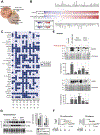
HCV-induced epigenetic changes persist after HCV clearance in patient-derived liver tissue. (A) Approach: HCV-induced H3K27ac histone modifications were measured genome-wide using a ChIPmentation-based ChIP-Seq protocol optimized for low input material such as patient-derived liver biopsy samples and resections. (B) Unsupervised clustering of normalized read counts in ChIP-Seq peaks of 12,700 genes linked with significant (P < .05) H3K27ac modifications in HCV-infected (n = 18), DAA-cured (n = 8), HBV-infected (n = 4), or NASH (n = 7) vs noninfected control (n = 6) patients.
Figure 2.
Figure 2.
HCV-infection induces specific epigenetic changes in the liver of HCV-infected patients. (A) PCA for control, noninfected, HCV-infected, DAA-cured, IFN-cured, HBV-infected, and NASH patient samples. Comparative analysis of epigenetic modifications separated based on fibrosis score along the primary component (dimension 1). (B) H3K27ac modifications among HCV-infected patients correlate (Spearman rank correlation coefficients and P values) with H3K27ac modifications among NASH or HBV-infected patients. Common H3K27ac modifications were analyzed. Prognostic association of hepatic gene expression was determined by using Cox score for time to overall death in a cohort of patients as previously described. (C) HCV-induced and persistent epigenetic changes after DAA cure in patient-derived liver tissue are associated with a decreased survival and death. H3K27ac modifications among HCV-infected correlate with persistent H3K27ac modifications among DAA-cured patients. (D) H3K27ac modifications correlate with significantly differentially expressed genes in HCV-infected and DAA-cured patients.
Figure 3.
Figure 3.
Pathway analysis of epigenetic and transcriptional reprogramming in HCV-infected patients unravels candidate genes driving carcinogenesis after DAA cure. (A) Hallmark pathways significantly enriched for H3K27ac modifications in infected (n = 18) or/and DAA-cured (n = 8) compared with control (n = 6) patient samples. A large overlap of enriched pathways persists in DAA-cured patients. (B) Venn diagram showing HCC risk gene candidates as the overlap of significantly modified genes in HCV-infected (F1–F4) and DAA-cured (F2–F3 and F4) patients derived from the ChIP-Seq experiment shown in Figure 1B. (C) Oncogenes (red) and TSGs (green) from the 2193 potential HCC risk gene candidates, with their biological functions indicated. (D) Heat map depicting transcriptional changes of the oncogenes and TSGs described in C in HCV-infected and DAA-cured patients. (E) Genes with persistent HCV-induced H3K27ac modifications after DAA cure, linked with the 32-gene prognostic liver signature predicting HCC in HCV-infected patients,, and overlapped with the hallmarks of cancer. Oncogenes shown in D are highlighted in black. This network includes 910 potential HCC risk gene candidates, highlighting a strong enrichment for modifications linked to carcinogenesis. EMT, epithelial–mesenchymal transition; IL2, interleukin 2; PI3K, phosphoinositide 3-kinase; STAT5, signal transducer and activator of transcription 5.
Figure 4.
Figure 4.
HCV-induced epigenetic changes persisting after DAA-based cure are present in the tumor tissue of patients with DAA-cured HCC. H3K27ac modifications from patient-derived resections of tumor and nontumor adjacent paired tissue samples. Similar to the analysis shown in Figure 1B, we performed an unsupervised clustering of normalized read counts in ChIP-Seq peaks of 7609 genes linked with significant (q < 0.05) H3K27ac modifications in DAA-cured adjacent (n = 8) or paired-tumor (n = 8) tissues vs noninfected control patients (n = 6). The proportions (percentages) of common (yellow) or distinct genes associated with changes in H3K27ac levels in tumor (blue) or nontumor paired-adjacent tissues (orange) are represented as a pie chart. N, nontumor; T, tumor.
Figure 5.
Figure 5.
Analysis of H3K27ac changes in livers of HCV-infected humanized mice identifies virus-specific modifications in human hepatocytes. (A) Our experimental setup: uPA-SCID mice were infected with HCV for 8 weeks and cured with a combination of DAAs MK7009 (50 or 100 mg/kg/d) and BMS-788329 (20 mg/kg/d) for 16 weeks. Livers were analyzed at week 24 by ChIP-Seq and RNA-Seq. Human albumin level (left) and HCV viral load (right) were measured to monitor functional engrafted human hepatocytes and HCV clearance after DAA treatment, respectively. (B) Unsupervised clustering of normalized read counts in ChIP-Seq peaks of 2483 genes linked with significant (q < 0.05) H3K27ac modifications in HCV-infected (n = 5) or DAA-cured (n = 5) vs noninfected control (n = 5) mice. (C) Hallmark pathways significantly enriched for H3K27ac modifications in infected (n = 5) or/and DAA-cured (n = 5) compared with noninfected (n = 5) mice samples. A significant overlap of enriched pathways persists in DAA-cured mice. (D) Venn diagram showing the HCV-induced and persistent genes with H3K27ac changes as the overlap of significantly modified genes in HCV-infected and DAA-cured mice derived from the ChIP-Seq experiment shown in B. (E) Oncogenes (red) and TSGs (green) with persistent HCV-induced H3K27ac modifications identified in the 306 HCV-induced and persistent genes with H3K27ac changes, with their biological functions indicated. (F) Heat map depicting transcriptional changes of the oncogenes and TSGs described in E in HCV-infected humanized and DAA-cured mice. EMT, epithelial–mesenchymal transition; NFκB, nuclear factor κB.
Figure 6.
Figure 6.
Intersection of ChIP-Seq and RNA-Seq analyses from livers of patients and humanized mice uncovers HCV-induced persistent epigenetic changes associated with HCC risk after SVR. (A) Venn diagram showing the overlap of H3K27ac modifications between the human HCC risk gene candidates and significantly modified genes in HCV-infected and DAA-cured mice derived from the ChIP-Seq experiments shown in Figures 1B and 5B, respectively. (B) Expression data of genes with significant H3K27ac changes from livers of HCV-infected and DAA-cured patients (n = 32) and mice (n = 15) were intersected to uncover common genes with HCV-induced and persistent epigenetic and transcriptional changes after DAA. (C) Presence of epigenetic modifications on the 38 identified genes in pairwise liver tissues from DAA-cured patients. H3K27ac modifications (vs control liver samples) were assessed on the corresponding genes in nontumorous adjacent and HCC liver tissues from DAA-cured patients. Dark blue squares represent increased H3K27ac changes and light blue squares represent unchanged status. (D) Analysis of protein level of SPHK1 and SOX9 protein in control, HCV-infected, and DAA-cured mice by western blot. (E) Analysis of SPHK1 and SOX9 protein levels in control (n = 7), HCV-infected (non-HCC and HCC; n = 8) and DAA-cured (non-HCC and HCC; n = 8) patients by western blot. One representative gel of 4 is shown. Graphs show quantification of western blot intensities in arbitrary units normalized to total protein level (Ponceau staining). Results show mean ± standard error of the mean of integrated blot densities. (F) Probability of HCC development according to the gene expression level of SPHK1 among 216 patients with HCV-induced cirrhosis or 21 patients with HCC occurrence after HCV cure.

Comment in

References

    1. Ryerson AB, Eheman CR, Altekruse SF, et al. Annual report to the nation on the status of cancer, 1975–2012, featuring the increasing incidence of liver cancer. Cancer 2016;122:1312–1337. - PMC - PubMed
    1. Fujiwara N, Friedman SL, Goossens N, Hoshida Y. Risk factors and prevention of hepatocellular carcinoma in the era of precision medicine. J Hepatol 2018;68:526–549. - PMC - PubMed
    1. Boldanova T, Suslov A, Heim MH, Necsulea A. Transcriptional response to hepatitis C virus infection and interferon-alpha treatment in the human liver. EMBO Mol Med 2017;9:816–834. - PMC - PubMed
    1. El-Serag HB, Kanwal F, Richardson P, Kramer J. Risk of hepatocellular carcinoma after sustained virological response in veterans with hepatitis C virus infection. Hepatology 2016;64:130–137. - PMC - PubMed
    1. Kanwal F, Kramer J, Asch SM, et al. Risk of hepatocellular cancer in HCV patients treated with direct-acting antiviral agents. Gastroenterology 2017;153:996–1005 e1. - PubMed

Publication types

MeSH terms

Substances