. 2019 Apr:76:99-107.
doi: 10.1016/j.dnarep.2019.02.008. Epub 2019 Feb 23.

RAD51D splice variants and cancer-associated mutations reveal XRCC2 interaction to be critical for homologous recombination

Affiliations

RAD51D splice variants and cancer-associated mutations reveal XRCC2 interaction to be critical for homologous recombination

Robert A Baldock et al. DNA Repair (Amst). 2019 Apr.

Abstract

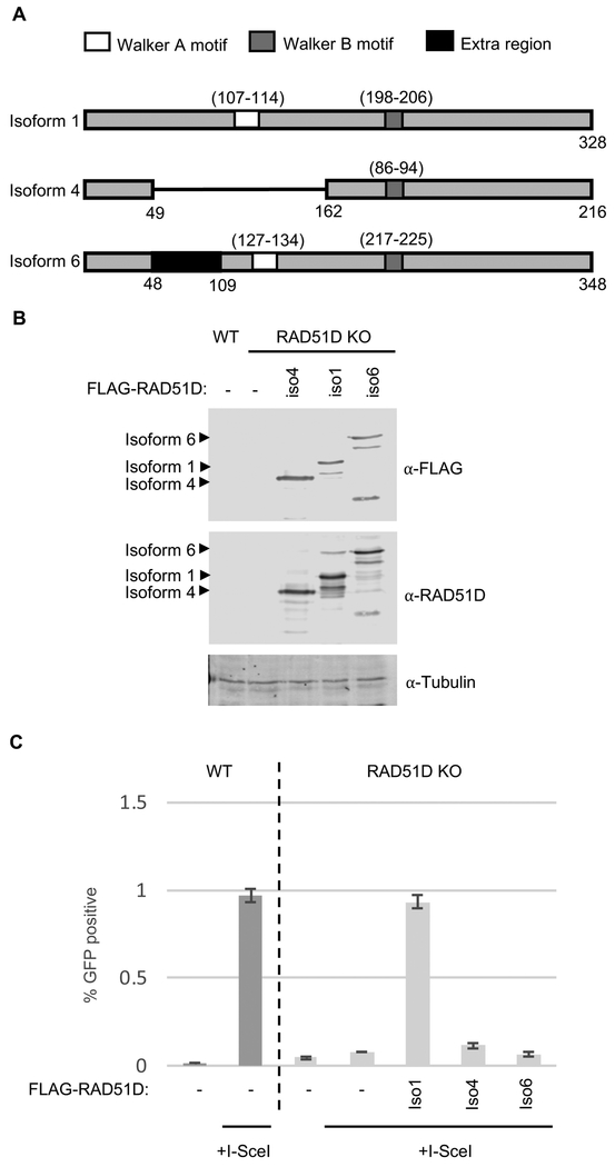
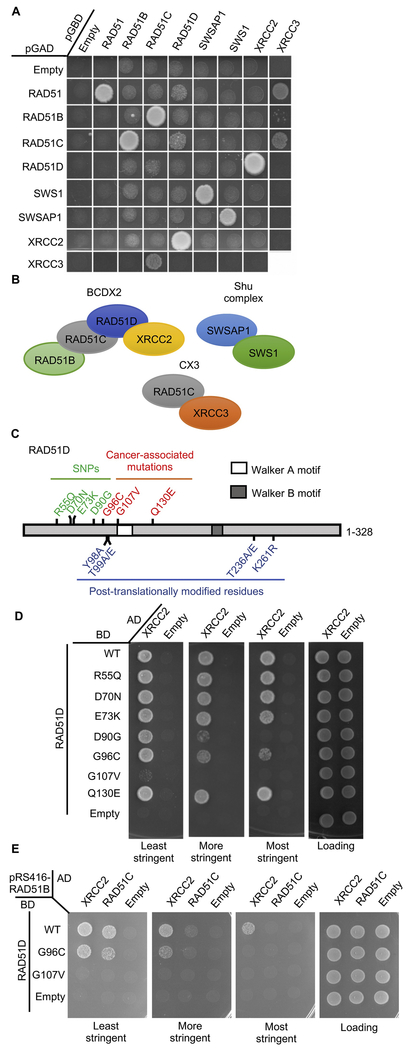
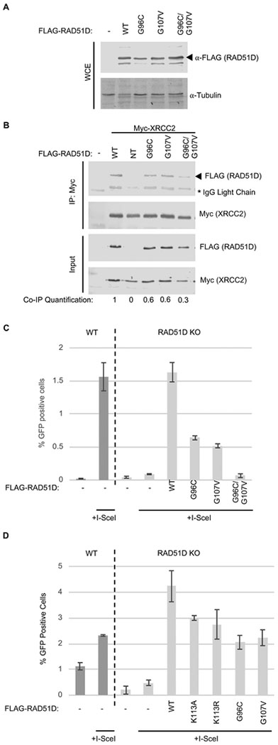
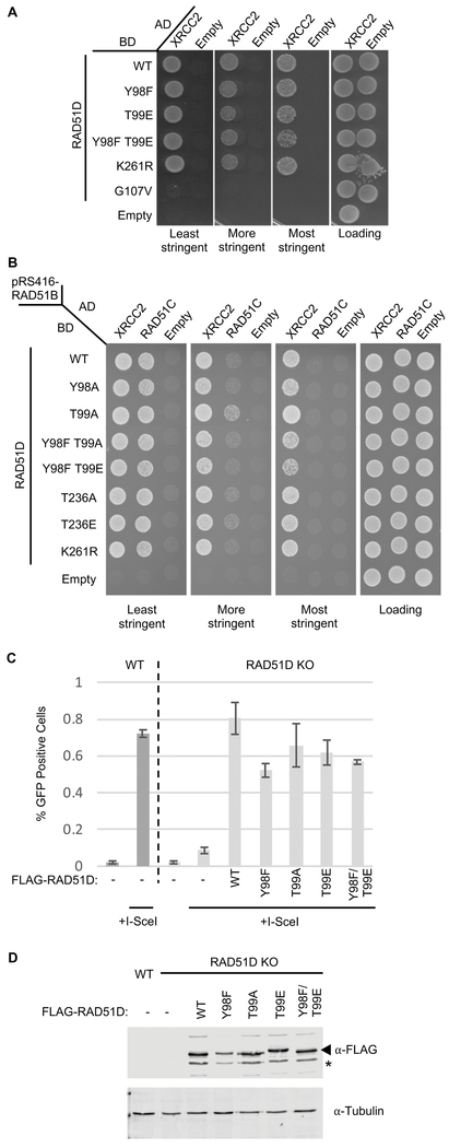
The proficiency of cancer cells to repair DNA double-strand breaks (DSBs) by homologous recombination (HR) is a key determinant in predicting response to targeted therapies such as PARP inhibitors. The RAD51 paralogs work as multimeric complexes and act downstream of BRCA1 to facilitate HR. Numerous epidemiological studies have linked RAD51 paralog mutations with hereditary cancer predisposition. Despite their substantial links to cancer, RAD51 paralog HR function has remained elusive. Here we identify isoform 1 as the functional isoform of RAD51D, whereas isoform 4 which has a large N-terminal deletion (including the Walker A motif), and isoform 6 which includes an alternate exon in the N-terminus, are non-functional. To determine the importance of this N-terminal region, we investigated the impact of cancer-associated mutations and SNPs in this variable RAD51D N-terminal region using yeast-2-hybrid and yeast-3-hybrid assays to screen for altered protein-protein interactions. We identified two cancer-associated mutations close to or within the Walker A motif (G96C and G107 V, respectively) that independently disrupt RAD51D interaction with XRCC2. We validated our yeast interaction data in human U2OS cells by co-immunoprecipitation and determined the impact of these mutations on HR-proficiency using a sister chromatid recombination reporter assay in a RAD51D knock-out cell line. Our investigation reveals that the interaction of RAD51D with XRCC2 is required for DSB repair. By characterizing the impact of cancer-associated mutations on RAD51D interactions, we aim to develop predictive models for therapeutic sensitivity and resistance in patients who harbor similar mutations in RAD51D.

Keywords: Double-strand break repair; Homologous recombination; RAD51 paralogs; RAD51D; Walker A motif; XRCC2.


Conflict of interest statement

Conflicts of interest

The authors declare that there are no conflicts of interest.

Figures

Figure 1
Figure 1. RAD51D splicing isoform 1, but not isoforms 4 or 6, rescues HR in RAD51D knock-out U20S cell lines.
A) Schematic diagram of the three coding isoforms of RAD51D (isoforms 1,4 and 6). Amino acid residue numbers of the Walker A (white box) and B (dark grey box) motifs are shown in parenthesis. The extra coding region in isoform 6 is shown in black. B) U20S wild-type or RAD51D knock-out cells were seeded on 35mm plates 24-hours prior to transfection with FLAG-RAD51D cDNA constructs. Following transfection, cells were incubated for an additional 24-hours before lysis and Western blot analysis for FLAG expression using α-Flag antibodies. α-Tubulin was used as a loading control. C) Cells were seeded 24-hours prior to transfection as in B. Cells were co-transfected with I-Scel and FLAG-RAD51D cDNAs or an empty vector control. Cells were incubated for an additional 48-hours and then analyzed by flow cytometry for green-fluorescence. The bar chart shows the average percentage of GFP+ cells over three experiments. Error bars show one standard deviation from the mean.
Figure 2
Figure 2. Identification of cancer-associated mutations, G96V and G107V, in RAD51D that impair its interaction with XRCC2 and RAD51C.
A) Yeast 2-hybrid analysis showing the interactions of each of the RAD51 paralogs (and SWS1, a SWIM-domain containing protein) with each other. The PJ694a yeast strain was transformed with a plasmid where RAD51, the RAD51 paralogs (RAD51B, RAD51C RAD51D, SWSAP1, XRCC2, and XRCC3) and SWS1 were cloned into a plasmid that expresses the GAL4 DNA activating (pGAD) and GAL4 DNA binding (pGBD) domains.A yeast-2-hybrid interaction was assayed by platting the yeast on SC-Leu¯Trp¯His¯ and compared to the empty pGAD or pGBD plasmids, which were used as negative controls. Note that XRCC3 analysis had to be performed on a separate plate because of the plate size and that, consistent with the yeast Rad51 paralogs, human RAD51 Y2H interactions with the RAD51 paralogs are only observed when RAD51 is expressed in the pGAD vector [57]. B) Diagram representing the protein-protein interaction between the RAD51 paralog subcomplexes shown in the part A. BCDX2 is composed of RAD51B (light green), RAD51C (grey), RAD51D (blue), and XRCC2 (yellow). The CX3 complex is composed of RAD51C (grey) and XRCC3 (orange) and the Shu complex composed of SWSAP1 (light blue) and SWS1 (green). C) Schematic diagram showing RAD51D Walker motifs (white and dark grey boxes) as well as SNPs (green), cancer-associated mutations (red) and potentially post-translationally modified residues (blue) analyzed. D) Yeast-2-hybrid analysis of SNPs and cancer-associated mutations in RAD51D on its interaction with XRCC2. The PJ694a yeast strain was transformed with a plasmid where RAD51D or the indicated RAD51D mutation was fused to the GAL4-DNA binding domain [BD; pGBD-RAD51D (WT)] and a plasmid where XRCC2 was fused to the GAL4-DNA activating domain (AD; pGAD-XRCC2). A yeast-2-hybrid interaction was assayed by platting the yeast on SC-Leu¯Trp¯His¯ (least stringent), SC-Leu¯Trp¯His¯+3AT (more stringent), or SC-Leu¯Trp¯His¯Ade¯ (most stringent) and compared to the loading control (SC-Leu¯Trp¯). Empty AD (pGAD; Empty) plasmid was used as a negative control. E) Yeast-3-hybrid analysis of RAD51D-G96C and/or -G107V mutations with XRCC2 and RAD51C. The PJ694a yeast strain was transformed with three plasmids; 1) a plasmid where RAD51D (or the indicated mutant) was fused to the GAL4-DNA binding domain [BD; pGBD-RAD51D (WT)], 2) a plasmid where either XRCC2 or RAD51C was fused to the GAL4-DNA activating domain (AD; pGAD-XRCC2, pGAD-RAD51C), and 3) a plasmid that constitutively expressed RAD51B (pRS416-RAD51B). Note that a plasmid containing RAD51B was used to help stabilize RAD51C expression. A yeast-3-hybrid interaction between RAD51D with XRCC2 or RAD51C was assayed by platting the yeast on SC-Leu¯Trp¯Ura¯His¯ (least stringent), SC-Leu¯Trp¯Ura¯His¯+3AT (more stringent), or SC-Leu¯Trp¯Ura¯His¯Ade¯ (most stringent) and compared to the loading control SC-Leu¯Trp¯Ura¯ (Loading). Empty pGBD (BD Empty) and pGAD (AD Empty) plasmids were used as negative controls.
Figure 3
Figure 3. RAD51D-G96C, -G107V and the double -G96C/G107V mutant exhibit impaired XRCC2 interaction and HR in human U20S cells.
A) U20S wild-type or RAD51D knock-out cells were seeded on 35mm plates 24-hours prior to transfection with FLAG-RAD51D mutant cDNA constructs. Following transfection, cells were incubated for a further 24-hours before lysis and Western blot analysis for FLAG expression using aFLAG antibodies. α-Tubulin was used as a loading control. B) Co-immunoprecipitation of Myc-XRCC2 with FLAG-RAD51D-G96C, G107V and the double-mutant from human U20S cells. U20S cells were seeded on 100mm plates 24-hours prior to transfection with FLAG-RAD51D wild-type or mutant cDNA constructs (NT= not transfected). Following transfection, cells were incubated for an additional 24-hours before lysis. Lysates were incubated overnight at 4°C with α-Myc agarose beads. The next day, the agarose beads were washed and the co-immunoprecipitates were eluted using SDS-sample buffer. Samples were analyzed by Western blotting for a- FLAG (co-immunoprecipitation) and α-Myc (immunoprecipitation). Western blot band densitometry was calculated using Image Studio software, co-immunoprecipitation FLAG-band intensity (Co-IP Quantification) was normalized to immunoprecipitation (IP: Myc) and to input FLAG expression to account for transfection efficiency and any variability in the immunoprecipitation. *Signifies the bands produced by cross-reactivity of the secondary antibody with IgG light-chain of the Myc antibody used for immunoprecipitation. C) U20S wild-type or RAD51D knock-out cells were seeded on 35mm plates 24-hours prior to transfection with the indicated FLAG-RAD51D cDNA mutant constructs. Cells were co-transfected with either l-Scel and FLAG-RAD51D cDNAs or l-Scel and an empty vector control. Cells were incubated for an additional 48-hours and analyzed by flow cytometry for green-fluorescence. The bar chart shows the average percentage of GFP+ cells over three experiments. Error bars show one standard deviation from the mean. D) U20S wild-type or RAD51D knock-out cells were seeded and transfected as in 3C (including K113A and K113R; Walker A motif mutants). Cells were incubated for an additional 48-hours and analyzed by flow cytometry for green-fluorescence. The bar chart shows the average percentage of GFP+ cells over three experiments. Error bars show one standard deviation from the mean
Figure 4
Figure 4. Mutations of residues in RAD51D that are potentially post-translationally modified do not affect the interaction of RAD51D with RAD51C or XRCC2 and do not affect HR-proficiency.
A) Yeast 2-hybrid analysis of potential post-translationally modified residues in RAD51D on its interaction with XRCC2. The PJ694a yeast strain was transformed with a plasmid where RAD51D or the indicated RAD51D mutation was fused to the GAL4-DNA binding domain [BD; pGBD-RAD51D (WT)] and a plasmid where XRCC2 was fused to the GAL4-DNA activating domain (AD; pGAD-XRCC2). A yeast-2-hybrid interaction was assayed by platting the yeast on SC-Leu¯Trp¯His¯ (least stringent), SC-Leu¯Trp¯His¯+3AT (more stringent), or SC-Leu¯Trp¯His¯Ade¯(most stringent) and compared to the loading control (SC-Leu¯Trp¯). Empty AD (pGAD; Empty) plasmid was used as a negative control. B) Yeast 3-hybrid analysis of potential post-translationally modified residues in RAD51D on its interaction with XRCC2 and RAD51C. The PJ694a yeast strain was transformed with three plasmids; 1) a plasmid where RAD51D (or the indicated mutant) was fused to the GAL4-DNA binding domain [BD; pGBD-RAD51D (WT)], 2) a plasmid where either XRCC2 or RAD51C was fused to the GAL4-DNA activating domain (AD; pGAD-XRCC2, pGAD-RAD51C), and 3) a plasmid that constitutively expressed RAD51B (pRS416- RAD51B). Note that a plasmid containing RAD51B was used to help stabilize RAD51C expression. A yeast-3-hybrid interaction between RAD51D with XRCC2 or RAD51C was assayed by platting the yeast on SC-Leu¯Trp¯Ura¯His¯ (least stringent), SC-Leu¯Trp¯Ura¯His¯+3AT (more stringent), or SC-Leu¯Trp¯Ura¯His¯Ade¯(most stringent) and compared to the loading control SC-Leu¯Trp¯Ura¯ (Loading). Empty pGBD (BD Empty) and pGAD (AD Empty) plasmids were used as negative controls. C) U20S wild-type (WT) or RAD51D knock-out cells were seeded on 35mm plates 24- hours prior to transfection with FLAG-RAD51D cDNA mutant constructs. Cells were cotransfected with either l-Scel and FLAG-RAD51D cDNAs containing residue substitutions or l-Scel and an empty vector control. Cells were incubated for an additional 48-hours after which, cells were analyzed by flow cytometry for green- fluorescence. The bar chart shows the average percentage of GFP+ cells over three experiments. Error bars show one standard deviation from the mean. D) U20S wild-type (WT) or RAD51D knock-out cells were seeded on 35mm plates 24- hours prior to transfection with FLAG-RAD51D cDNA containing residue substitutions. Following transfection, cells were incubated for an additional 24-hours before lysis and Western blot analysis for RAD51D expression was done using αFLAG and αRAD51D antibodies. α-Tubulin was used as a loading control. The star indicates a non-specific band.

References

    1. Jasin M and Rothstein R, Repair of strand breaks by homologous recombination. Cold Spring Harb Perspect Biol, 2013. 5(11): p. a012740. - PMC - PubMed
    1. Renkawitz J, Lademann CA, and Jentsch S, Mechanisms and principles of homology search during recombination. Nat Rev Mol Cell Biol, 2014. 15(6): p. 369–383. - PubMed
    1. Chun J, Buechelmaier ES, and Powell SN, Rad51 paralog complexes BCDX2 and CX3 act at different stages in the BRCA1-BRCA2-dependent homologous recombination pathway. Mol Cell Biol, 2013. 33(2): p. 387–95. - PMC - PubMed
    1. Liu T, et al., hSWS1.SWSAP1 is an evolutionarily conserved complex required for efficient homologous recombination repair. J Biol Chem, 2011. 286(48): p. 41758–66. - PMC - PubMed
    1. Godin SK, Sullivan MR, and Bernstein KA, Novel insights into RAD51 activity and regulation during homologous recombination and DNA replication. Biochemistry and Cell Biology, 2016: p. 1–12. - PMC - PubMed

Publication types