. 2019 Apr;568(7751):244-248.
doi: 10.1038/s41586-019-1027-4. Epub 2019 Mar 5.

HIV-1 remission following CCR5Δ32/Δ32 haematopoietic stem-cell transplantation

Affiliations

HIV-1 remission following CCR5Δ32/Δ32 haematopoietic stem-cell transplantation

Ravindra K Gupta et al. Nature. 2019 Apr.

Abstract

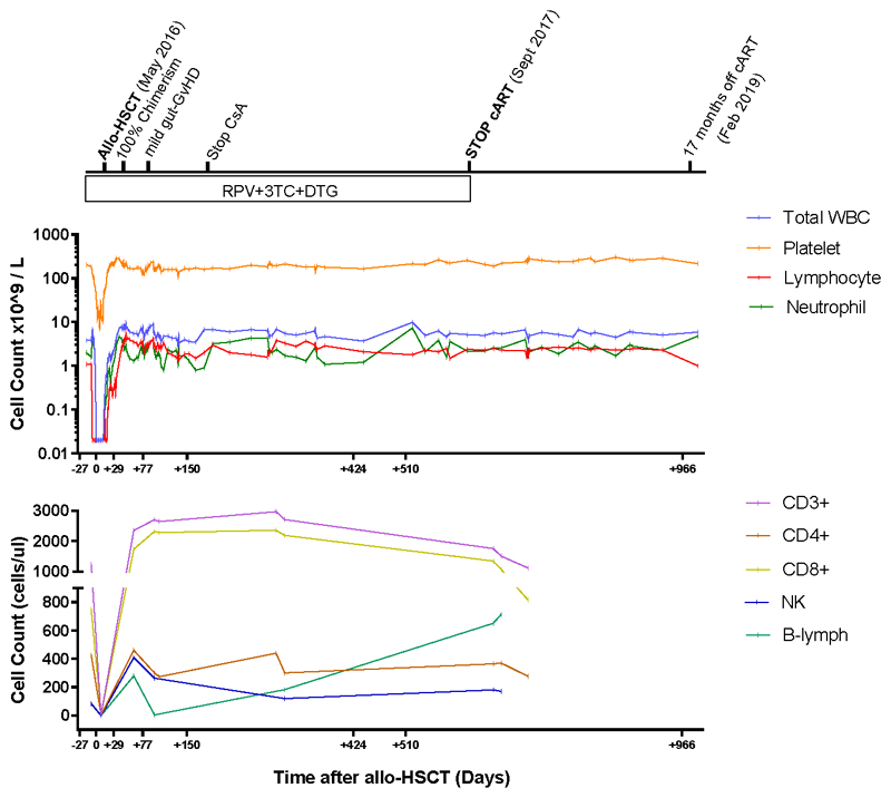
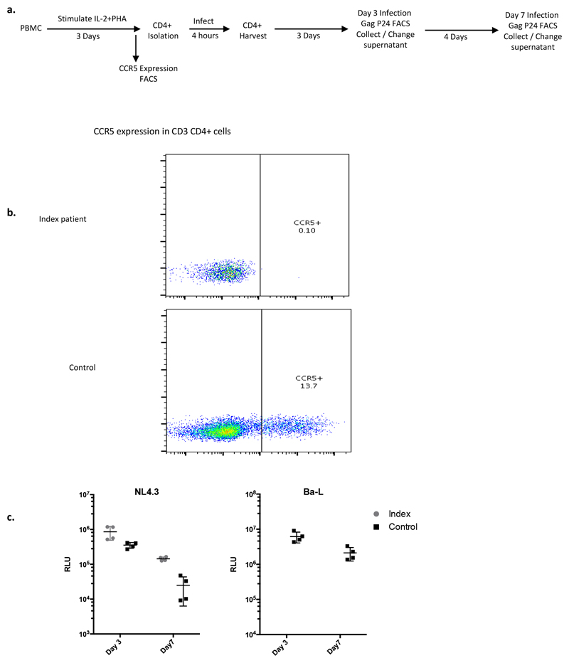
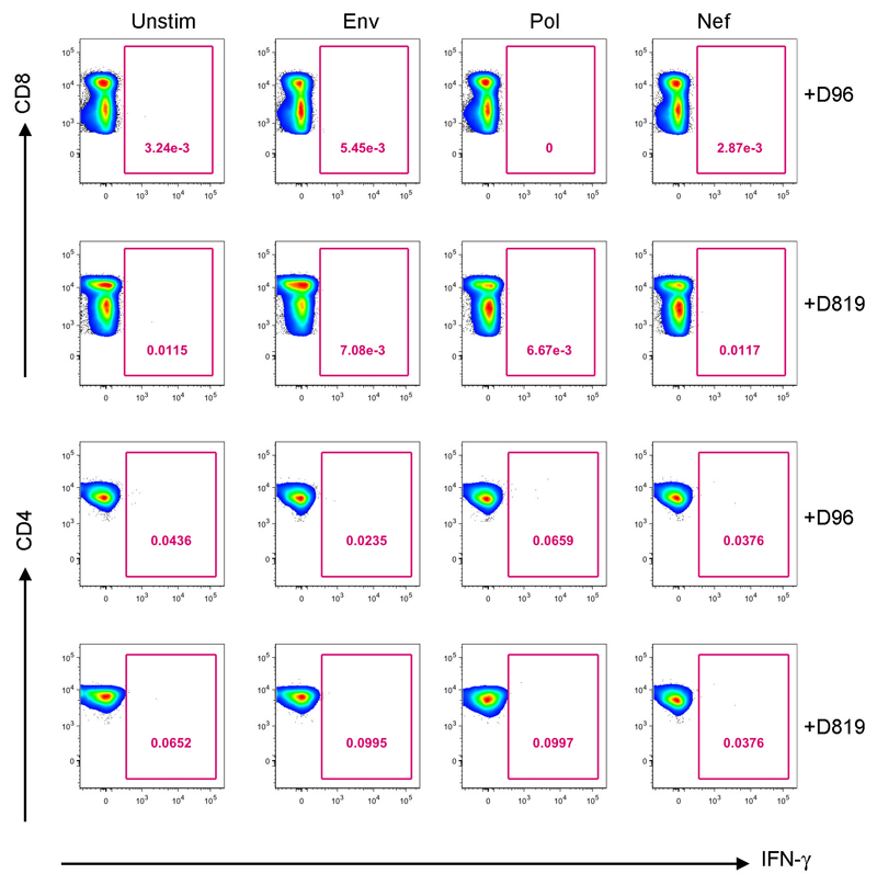
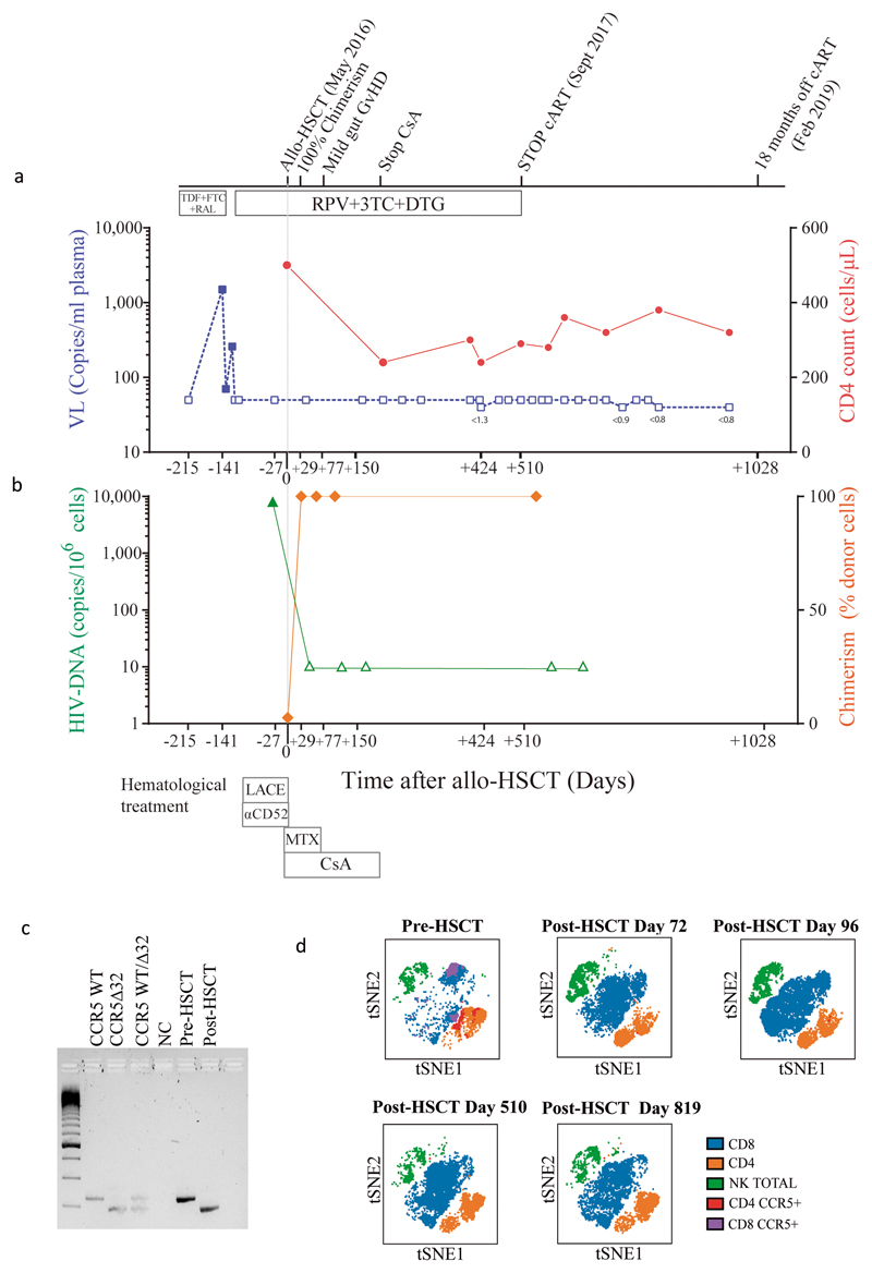
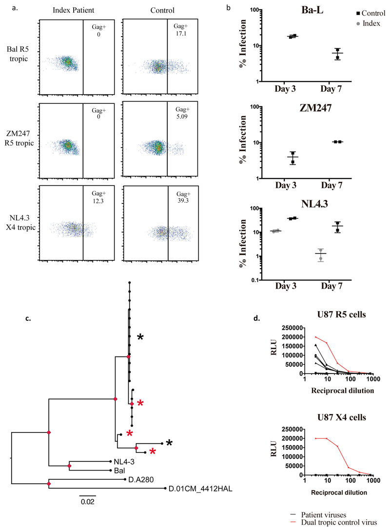
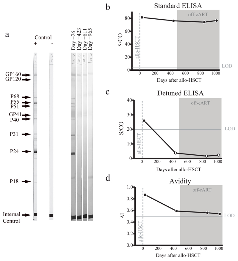
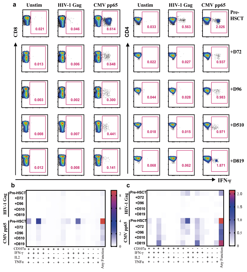
A cure for HIV-1 remains unattainable as only one case has been reported, a decade ago1,2. The individual-who is known as the 'Berlin patient'-underwent two allogeneic haematopoietic stem-cell transplantation (HSCT) procedures using a donor with a homozygous mutation in the HIV coreceptor CCR5 (CCR5Δ32/Δ32) to treat his acute myeloid leukaemia. Total body irradiation was given with each HSCT. Notably, it is unclear which treatment or patient parameters contributed to this case of long-term HIV remission. Here we show that HIV-1 remission may be possible with a less aggressive and toxic approach. An adult infected with HIV-1 underwent allogeneic HSCT for Hodgkin's lymphoma using cells from a CCR5Δ32/Δ32 donor. He experienced mild gut graft-versus-host disease. Antiretroviral therapy was interrupted 16 months after transplantation. HIV-1 remission has been maintained over a further 18 months. Plasma HIV-1 RNA has been undetectable at less than one copy per millilitre along with undetectable HIV-1 DNA in peripheral CD4 T lymphocytes. Quantitative viral outgrowth assays from peripheral CD4 T lymphocytes show no reactivatable virus using a total of 24 million resting CD4 T cells. CCR5-tropic, but not CXCR4-tropic, viruses were identified in HIV-1 DNA from CD4 T cells of the patient before the transplant. CD4 T cells isolated from peripheral blood after transplantation did not express CCR5 and were susceptible only to CXCR4-tropic virus ex vivo. HIV-1 Gag-specific CD4 and CD8 T cell responses were lost after transplantation, whereas cytomegalovirus-specific responses were detectable. Similarly, HIV-1-specific antibodies and avidities fell to levels comparable to those in the Berlin patient following transplantation. Although at 18 months after the interruption of treatment it is premature to conclude that this patient has been cured, these data suggest that a single allogeneic HSCT with homozygous CCR5Δ32 donor cells may be sufficient to achieve HIV-1 remission with reduced intensity conditioning and no irradiation, and the findings provide further support for the development of HIV-1 remission strategies based on preventing CCR5 expression.


Conflict of interest statement

Competing interests

The authors have no conflicts of interest

Figures

Extended data figure 1
Extended data figure 1. Blood Cell populations over time.
Abbreviations: HSCT: haematopoietic stem cell transplantation; CsA ciclosporin A; ART antiretroviral therapy; RPV rilpivirine; DTG dolutegravir; 3TC lamivudine
Extended data figure 2
Extended data figure 2. Susceptibility of index patient CD4 T cells to R5 tropic and X4 tropic HIV-1
a. Experimental flow for measurement of infection by intracellular p24 gag staining. Control cells were from a healthy HIV- CCR5+ donor. b. Flow cytometry analysis of PBMC following 3 days of stimulation exhibiting the expression pattern of CCR5 receptor within CD3+ CD4+ T cells in both healthy donor (control) and index patient. c. Culture supernatants from CD4 T cells infected with R5 and X4 tropic viruses were collected on days 3 and 7 to measure infectivity on HeLa TZM-bl reporter cells. Infectivity is measured as a reduction in Tat-induced firefly luciferase reporter gene expression in TZM-bl. Error bars represent standard error of the mean. N=2: one donor and one index patient Experiments were repeated 3 times with similar results.
Extended data figure 3
Extended data figure 3. CD8+T cell responses and CD4 T cell responses to HIV.
Representative FACS plots showing percentage of virus specific CD8+ T cells (top panel) and CD4+ T cells (bottom panel) identified via intracellular staining for IFN-g, following stimulation with HIV Pol, Env and Nef peptide pools post-HSCT at days +D96 and +D819.
Figure 1
Figure 1. Clinical course before and after allogeneic Hematological Stem Cell Transplantation
a. Antiretroviral treatment and chemotherapy/immunosuppression associated with allogeneic HSCT along with plasma viral load (HIV-1 RNA) and CD4 count over time. Small numbers below blue data points indicate results of ultra sensitive viral load assay. b. HIV-1 DNA in PBMC and donor chimerism in T cell fraction c. Genotyping of CCR5 alleles with agarose gel electrophoresis of PCR amplified DNA fragments using a 100 base pair DNA ladder; NC negative control. d. tSNE plots of PBMC pre and post HSCT showing CCR5 expression changes and cell population changes over time. Abbreviations: HSCT: haematopoietic stem cell transplantation LACE: lomustine Ara-C cyclophosphamide etoposide; MTX methotrexate; CsA ciclosporin A; ART antiretroviral therapy; RPV rilpivirine; DTG dolutegravir; 3TC lamivudine; RAL raltegravir; TDF tenofovir disoproxil fumarate; FTC emtricitabine. These experiments were carried out once only (a-d) and sample size is n=1 for all panels.
Figure 2
Figure 2. Susceptibility of index patient CD4 T cells to CCR5 tropic and CXCR4 tropic HIV-1 and coreceptor usage by index patient viruses prior to HSCT.
a. Representative plots of intracellular p24 gag staining within CD4+ T cell populations three days post infection of isolated CD4+ cells by CCR5 (R5) tropic viruses Bal and ZM247 and CXCR4 (X4) tropic virus NL4.3 b. Percentage infection in CD3+ CD4+ T cells determined by p24 staining at day 3 and day 7 post infection using R5 and X4 tropic viruses. Error bars represent standard error of the mean. Data are representative of 3 independent experiments each conducted in duplicate. c. Maximum likelihood phylogenetic tree showing single genome env C2-V5 sequences (HXB2 env 367-1533 nt) from PBMC prior to HSCT. NL4.3, Ba-L and two subtype D sequences from Genbank were also used. Red nodes indicate >70% bootstrap support. Gp120 amplicons from sequences marked with an asterix were cloned into a clade B gp160 Env plasmid. Virus env sequences marked by a red asterix generated infectious virus particles when co-transfected into HEK293T cells with the envelope deficient full-length HIV plasmid encoding luciferase (NL4-3 delta Env Luc). Black asterix indicates a sequence that did not generate infectious virus. d. Pseudoviruses from c. were used to infect U87 cells expressing either CXCR4 (X4) or CCR5 (R5). A dual tropic pseudovirus (WEAU-d15.410.787) was produced in parallel as a positive contro l for infection (red line). Experiments in a, b, c and d. were performed independently three times with similar results (sample size n=1 healthy control donor and n=1 index patient for a and b; sample size n=9 clones tested in d.).
Figure 3
Figure 3. HIV Specific Antibodies.
Humoral response dynamics were tested at days +26, +423, +811 and +965 after HSCT (last two time points in absence of ART). Antibody levels were measured using Western blot (a), the standard HIV-1 VITROS assay (b), a detuned low sensitive (LS) version of the HIV-1 VITROS assay (c), and the Limiting Antigen avidity assay (d). Open symbols represent values under the limit of detection. AI: Avidity Index; S/CO: Ratio signal/cut off; Allo-HSCT: allogeneic hematopoietic stem cell transplantation; ART: antiretroviral therapy. a, c, d were repeated twice independently with similar results.
Figure 4
Figure 4. HIV-Gag specific and CMV specific T cell responses.
a. Representative FACS plots showing percentage of virus specific CD8+ T cells (left panel) and CD4+ T cells (right panel) identified via intracellular staining for IFN-g, following stimulation with HIV-1 Gag or CMV pp65 peptide pools pre and post-HSCT at indicated days (+D72, +D96, +D510, +D819). A negative control containing PBMC from the same subject but without peptide mix was included (unstim) for each assay. b. Heat maps of levels of expression of CD107a, IFN-g, IL-2 and TNF-a in CD8+ T cells and (C) CD4+ T cells in response to HIV-1 Gag and CMV pp65 peptide stimulation pre and post-HSCT at indicated days subsequent to Boolean gating. Functions are listed beneath the heat maps with each of their respective combinations or any function detected. a. Experiment performed once due to limited cell numbers.

Comment in

References

    1. Hutter G, et al. Long-term control of HIV by CCR5 Delta32/Delta32 stem-cell transplantation. N Engl J Med. 2009;360:692–698. doi: 10.1056/NEJMoa0802905. - DOI - PubMed
    1. Allers K, et al. Evidence for the cure of HIV infection by CCR5Delta32/Delta32 stem cell transplantation. Blood. 2011;117:2791–2799. doi: 10.1182/blood-2010-09-309591. - DOI - PubMed
    1. UNAIDS. UNAIDS DATA. 2017 < http://www.unaids.org/en/resources/documents/2017/20170720_Data_book_2017>.
    1. Gupta RK, et al. HIV-1 drug resistance before initiation or re-initiation of first-line antiretroviral therapy in low-income and middle-income countries: a systematic review and meta-regression analysis. Lancet Infect Dis. 2018;18:346–355. doi: 10.1016/S1473-3099(17)30702-8. - DOI - PMC - PubMed
    1. TenoRes Study G. Global epidemiology of drug resistance after failure of WHO recommended first-line regimens for adult HIV-1 infection: a multicentre retrospective cohort study. Lancet Infect Dis. 2016;16:565–575. doi: 10.1016/S1473-3099(15)00536-8. - DOI - PMC - PubMed

Publication types

MeSH terms