. 2019 Mar 4;20(5):1103.
doi: 10.3390/ijms20051103.

Salidroside Ameliorates Renal Interstitial Fibrosis by Inhibiting the TLR4/NF-κB and MAPK Signaling Pathways

Affiliations

Salidroside Ameliorates Renal Interstitial Fibrosis by Inhibiting the TLR4/NF-κB and MAPK Signaling Pathways

Rui Li et al. Int J Mol Sci. .

Abstract

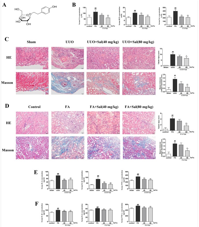
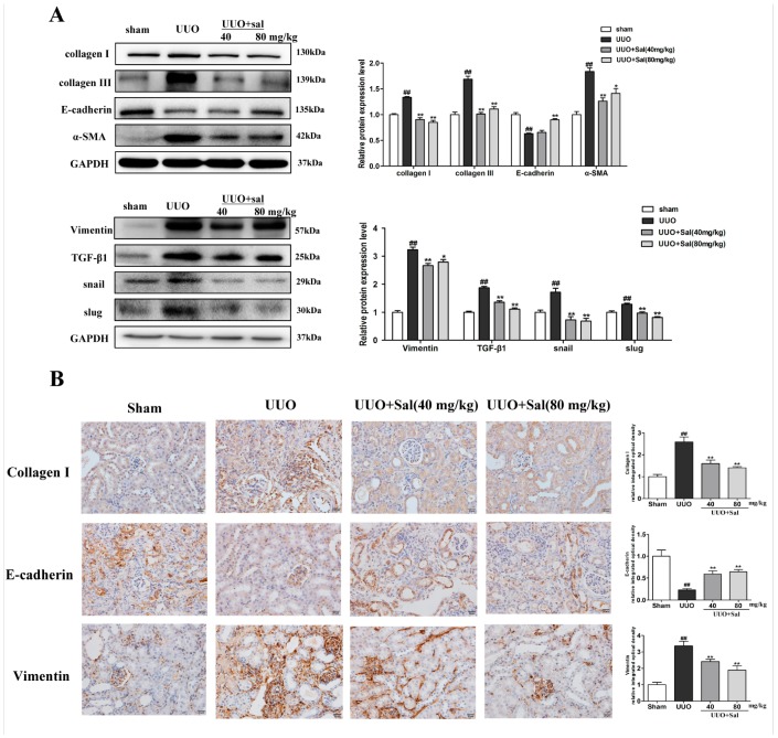
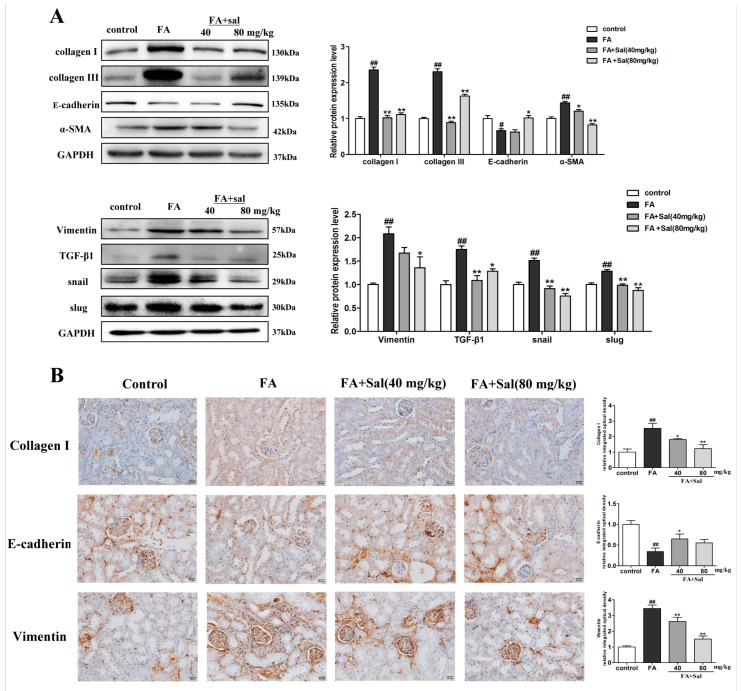
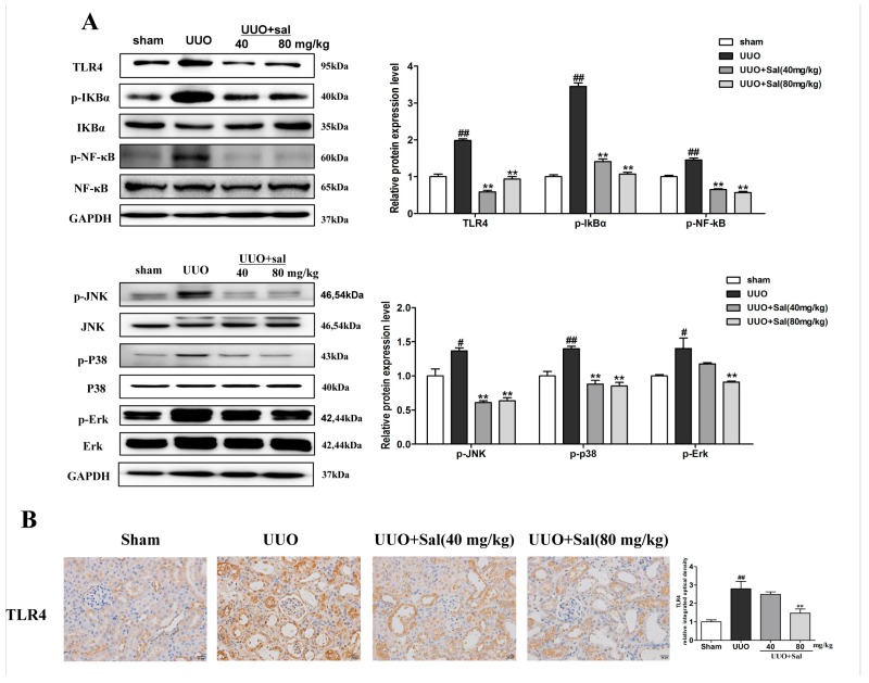
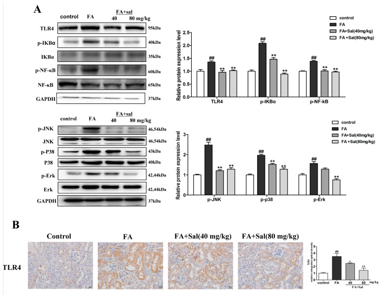
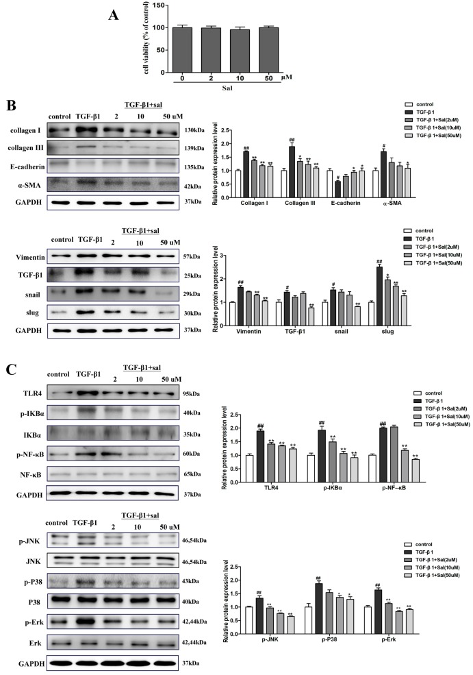
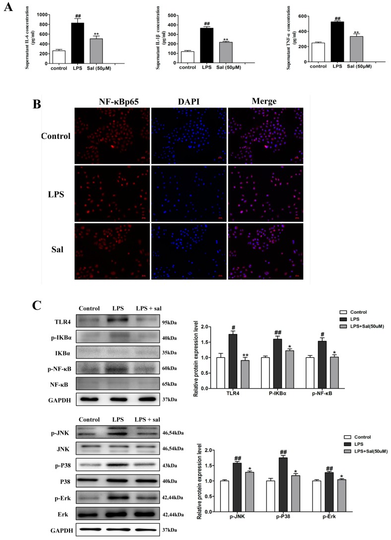
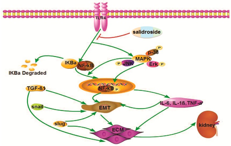
Salidroside (Sal) is an active ingredient that is isolated from Rhodiola rosea, which has been reported to have anti-inflammatory activities and a renal protective effect. However, the role of Sal on renal fibrosis has not yet been elucidated. Here, the purpose of the current study is to test the protective effects of Sal against renal interstitial fibrosis (RIF), and to explore the underlying mechanisms using both in vivo and in vitro models. In this study, we establish the unilateral ureteric obstruction (UUO) or folic acid (FA)-induced mice renal interstitial fibrosis in vivo and the transforming growth factor (TGF)-β1-stimulated human proximal tubular epithelial cell (HK-2) model in vitro. The levels of kidney functional parameters and inflammatory cytokines in serum are examined. The degree of renal damage and fibrosis is determined by histological assessment. Immunohistochemistry and western blotting are used to determine the mechanisms of Sal against RIF. Our results show that treatment with Sal can ameliorate tubular injury and deposition of the extracellular matrix (ECM) components (including collagen Ш and collagen I). Furthermore, Sal administration significantly suppresses epithelial-mesenchymal transition (EMT), as evidenced by a decreased expression of α-SMA, vimentin, TGF-β1, snail, slug, and a largely restored expression of E-cadherin. Additionally, Sal also reduces the levels of serum biochemical markers (serum creatinine, Scr; blood urea nitrogen, BUN; and uric acid, UA) and decreases the release of inflammatory cytokines (IL-1β, IL-6, TNF-α). Further study revealed that the effect of Sal on renal interstitial fibrosis is associated with the lower expression of TLR4, p-IκBα, p-NF-κB and mitogen-activated protein kinases (MAPK), both in vivo and in vitro. In conclusion, Sal treatment improves kidney function, ameliorates the deposition of the ECM components and relieves the protein levels of EMT markers in mouse kidneys and HK-2 cells. Furthermore, Sal treatment significantly decreases the release of inflammatory cytokines and inhibits the TLR4/NF-κB and MAPK signaling pathways. Collectively, these results suggest that the administration of Sal could be a novel therapeutic strategy in treating renal fibrosis.

Keywords: TLR4; epithelial-mesenchymal transition; inflammation; renal interstitial fibrosis; salidroside.


Conflict of interest statement

The authors declare no conflict of interest.

Figures

Figure 1
Figure 1
Sal alleviated renal function parameters, histopathology and inflammation in renal fibrotic mice. (A) The structure of salidroside (Sal). (B) Sal decreases the serum levels of serum creatinine (Scr), blood urea nitrogen (BUN) and uric acid (UA) in FA mice (n = 8). (C) Representative images of hematoxylin and eosin (H&E) and Masson staining of kidney tissue in unilateral ureteric obstruction (UUO) mice (n = 3). Original magnification: ×200. (D) Representative images of H&E and Masson staining of kidney tissue in folic acid (FA) mice (n = 3). The score of the lesion was determined to be either 0.5 points (slight or very little), 1 point (mild or small), 2 points (moderate or more), 3 points (severe or more quantity), or four points (extremely severe or a lot). When there was no obvious lesion, the score was 0 points. Original magnification: ×200. (E) The serum levels of TNF-α, IL-1β, and IL-6 in UUO mice were determined using enzyme-linked immunosorbent assay (ELISA) kits (n = 6). (F) The serum levels of TNF-α, IL-1β, and IL-6 in FA mice were determined using ELISA kits (n = 6). All data are represented as mean ± SD. # P < 0.05, ## P < 0.01 vs. Sham group, * P < 0.05, ** P < 0.01 vs. UUO group; # P < 0.05, ## P < 0.01 vs. control group, * P < 0.05, ** P < 0.01 vs. FA group.
Figure 2
Figure 2
Sal reduced the accumulation of extracellular matrix components and blocked EMT in UUO mice. (A) Western blotting was performed to determine the protein expression of collagen I, collagen Ш, α-SMA, vimentin, E-cadherin, TGF-β1, snail, and slug in the kidney tissue of UUO mice (n = 3). (B) Immunochemical staining of collagen I, E-cadherin, and vimentin in the kidney tissue of UUO mice (n = 3). Original magnification: ×400. All data are represented as mean ± SD. # P < 0.05, ## P < 0.01 vs. Sham group, * P < 0.05, ** P < 0.01 vs. UUO group.
Figure 3
Figure 3
Sal reduced the accumulation of extracellular matrix (ECM) components and blocked EMT in FA mice. (A) Western blotting was performed to determine the protein expression levels of collagen I, collagen Ш, α-SMA, vimentin, E-cadherin, TGF-β1, snail, and slug in the kidney tissue of FA mice (n = 3). (B) Immunochemical staining of collagen I, E-cadherin and vimentin in the kidney tissue of FA mice (n = 3). Original magnification: ×200. All data are represented as mean ± SD. # P < 0.05, ## P < 0.01 vs. control group, * P < 0.05, ** P < 0.01 vs. FA group.
Figure 4
Figure 4
Sal suppresses renal inflammation via the TLR4/NF-κB and MAPK signaling pathways in UUO mice. (A) Western blot analysis of TLR4, p-IкBα, p-NF-κB, p-Ρ38, p-ERK, and p-JNK protein expression in the kidney tissue of UUO mice (n = 3). (B) Immunochemical staining of TLR4 in the kidney tissue of UUO mice (n = 3). Original magnification: ×200. All data are represented as mean ± SD. # P < 0.05, ## P < 0.01 vs. Sham group, * P < 0.05, ** P < 0.01 vs. UUO group.
Figure 5
Figure 5
Sal suppresses renal inflammation via the TLR4/NF-κB and MAPK signaling pathways in FA mice. (A) Western blot analysis of TLR4, p-IкBα, p-NF-κB, p-Ρ38, p-ERK, and p-JNK protein expression in the kidney tissue of FA mice (n = 3). (B) Immunochemical staining of TLR4 in the kidney tissue of FA mice (n = 3). Original magnification: ×200. All data are represented as mean ± SD. # P < 0.05, ## P < 0.01 vs. control group, * P < 0.05, ** P < 0.01 vs. FA group.
Figure 6
Figure 6
Sal inhibited TGF-β1-induced HK-2 cells ECM deposition, EMT and inflammatory response via the TLR4/NF-κB and MAPK signaling pathways. (A) Effect of different doses of salidroside on HK-2 cell viability. HK-2 cells (1 × 10⁴ cells/well) were exposed to a series concentration of salidroside (0, 2, 10, 50 μM) for 24 h. The viability of HK-2 cells was assessed by a cell counting kit-8 (CCK-8) assay. (B) Western blotting was performed to determine the protein expression levels of collagen I, collagen Ш, α-SMA, vimentin, E-cadherin, TGF-β1, snail, and slug in TGF-β1-induced HK-2 cells. The cell was incubated with different concentrations of Sal (2, 10, 50 μM), followed by stimulation with 5 ng/mL of TGF-β1 for 48 h. (C) Sal inhibited the expression of TLR4, p-IкBα, p-NF-κB, p-Ρ38, p-ERK, and p-JNK in TGF-β1-induced HK-2 cells. The cell was incubated with different concentrations of salidroside (2, 10, 50 μM), followed by stimulation with 5 ng/mL of TGF-β1 for 48 h. All data are expressed as mean ± SD, # P < 0.05, ## P < 0.01 vs. control group, * P < 0.05, ** P < 0.01 vs. TGF-β1 group.
Figure 7
Figure 7
Sal suppressed lipopolysaccharide (LPS)-induced inflammatory response. (A) The concentrations of IL-1β, IL-6, and TNF-α in the culture supernatant of HK-2 cells were detected by an enzyme-linked immunosorbent assay (ELISA) (n = 6). The cell was incubated with Sal (50 μM) and followed by stimulation with 1 ug/mL of LPS for 24 h. (B) Sal significantly suppresses the nuclear transport process of NF-κBp65 in LPS-induced HK-2 cells, as determined by immunofluorescence (n = 3). Original magnification: ×200. The cell was incubated with salidroside (50 μM) for 24 h. Then, LPS (1 μg/mL) stimulated the cell for 60 min. (C) Sal inhibited the LPS-induced increase in TLR4, p-IкBα, p-NF-κB, p-Ρ38, p-ERK, and p-JNK in the HK-2 cells, as determined by western blotting (n = 3). The cell was incubated with Sal (50 μM), followed by stimulation with 1 ug/mL of LPS for 24 h. All data are expressed as mean ± SD, # P < 0.01. ## P < 0.01 vs. control group, * P < 0.05, ** P < 0.01 vs. LPS group.
Figure 8
Figure 8
Schematic illustration of the role and mechanism of Sal when ameliorating renal fibrosis. Sal suppresses the TLR4/MAPK/NF-кB signaling pathways to inhibit the inflammatory response and EMT process.

Similar articles

Cited by

References

    1. Jha V., Garciagarcia G., Iseki K., Li Z., Naicker S., Plattner B., Saran R., Wang A.Y., Yang C.W. Chronic kidney disease: global dimension and perspectives. Lancet. 2013;382:260–272. doi: 10.1016/S0140-6736(13)60687-X. - DOI - PubMed
    1. Kohan D.E., Barton M. Endothelin and endothelin antagonists in chronic kidney disease. Kidney Int. 2014;86:896–904. doi: 10.1038/ki.2014.143. - DOI - PMC - PubMed
    1. Declèves A.E., Sharma K. Novel targets of antifibrotic and anti-inflammatory treatment in CKD. Nat. Rev. Nephrol. 2014;10:257–267. doi: 10.1038/nrneph.2014.31. - DOI - PMC - PubMed
    1. Klahr S., Morrissey J. Obstructive nephropathy and renal fibrosis. Am. J. Physiol. Renal. Physiol. 2002;283:861–875. doi: 10.1152/ajprenal.00362.2001. - DOI - PubMed
    1. Bohle A., Christ H., Grund K.E., Mackensen S. The Role of the Interstitium of the Renal Cortex in Renal Disease. Contrib. Nephrol. 1979;16:109. - PubMed

MeSH terms