. 2019 Mar 30;12(1):31-42.
doi: 10.15283/ijsc18084.

Alteration of Genomic Imprinting Status of Human Parthenogenetic Induced Pluripotent Stem Cells during Neural Lineage Differentiation

Affiliations

Alteration of Genomic Imprinting Status of Human Parthenogenetic Induced Pluripotent Stem Cells during Neural Lineage Differentiation

Hye Jeong Lee et al. Int J Stem Cells. .

Abstract

Background and objectives: Genomic imprinting modulates growth and development in mammals and is associated with genetic disorders. Although uniparental embryonic stem cells have been used to study genomic imprinting, there is an ethical issue associated with the destruction of human embryos. In this study, to investigate the genomic imprinting status in human neurodevelopment, we used human uniparental induced pluripotent stem cells (iPSCs) that possessed only maternal alleles and differentiated into neural cell lineages.

Methods: Human somatic iPSCs (hSiPSCs) and human parthenogenetic iPSCs (hPgiPSCs) were differentiated into neural stem cells (NSCs) and named hSi-NSCs and hPgi-NSCs respectively. DNA methylation and gene expression of imprinted genes related neurodevelopment was analyzed during reprogramming and neural lineage differentiation.

Results: The DNA methylation and expression of imprinted genes were altered or maintained after differentiation into NSCs. The imprinting status in NSCs were maintained after terminal differentiation into neurons and astrocytes. In contrast, gene expression was differentially presented in a cell type-specific manner.

Conclusions: This study suggests that genomic imprinting should be determined in each neural cell type because the genomic imprinting status can differ in a cell type-specific manner. In addition, the in vitro model established in this study would be useful for verifying the epigenetic alteration of imprinted genes which can be differentially changed during neurodevelopment in human and for screening novel imprinted genes related to neurodevelopment. Moreover, the confirmed genomic imprinting status could be used to find out an abnormal genomic imprinting status of imprinted genes related with neurogenetic disorders according to uniparental genotypes.

Keywords: Genomic imprinting; Induced-pluripotent stem cells; Neural stem cells; Parthenogenetic cells; in vitro model.


Conflict of interest statement

Potential Conflict of Interest

The authors indicate no potential conflicts of interest.

Figures
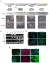
Fig. 1
Fig. 1
Derivation of NSCs from iPSCs and their characterization. (A) Schematic representation and phase contrast photomicrographs of hSi-NSCs and hPgi-NSCs derivation. hSi-NSCs, human somatic induced pluripotent stem cell-derived neural stem cells; hPgi-NSCs, human parthenogenetic induced pluripotent stem cell-derived neural stem cells. (B) RT-PCR analysis of the expression of pluripotency markers (OCT4 and NANOG), and self-renewal and NSC-specific markers (SOX2, SOX1, PAX6, and MASH1) in S Fibs, Pg Fibs, hSiPSCs, hPgiPSCs, and in differentiated hSi-NSCs and hPgi-NSCs. S Fibs, human somatic fibroblasts; Pg Fibs, human parthenogenetic fibroblasts; hSiPSCs, human somatic induced pluripotent stem cells; hPgiPSCs, human parthenogenetic induced pluripotent stem cells. (C) Immunocytochemistry of hSi-NSCs and hPgi-NSCs shows the expression of NSC marker proteins (SOX2 and PAX6; green), which co-localize with DAPI (blue) in the nucleus. Scale bar: 100 μm. (D) Immunocytochemistry of neurons and astrocytes differentiated from hSi-NSCs and hPgi-NSCs shows the expression of neuron marker proteins (MAP2, green; TUJ1, red), and an astrocyte marker protein (GFAP, green), which co-localize with DAPI (blue) in the nucleus. Scale bars: 100 μm. MAP2: microtubule-associated protein 2, TUJ1: neuron-specific class III beta-tubulin, GFAP: glial fibrillary acidic protein.
Fig. 2
Fig. 2
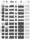
DNA methylation patterns of imprinted genes. Methylation of imprinted genes was analyzed by bisulfite sequencing. Paternally imprinted gene, MEG3; Maternally imprinted genes, MAGEL2, SNRPN, NDN and GRB10; the imprinted gene which have unmethylated CpGs, UBE3A. White and black circles indicate unmethylated and methylated CpG islands, respectively.
Fig. 3
Fig. 3
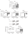
Expression of MEG3, MAGEL2, SNRPN, NDN, GRB10 (un1), and GRB10 (un2) analyzed using quantitative real-time PCR. The expression levels in hSiPSCs were set to 1. Means±SEM are shown for three independent experiments. Significance of inter-group differences was determined by one-way analysis of variance; #p<0.05, ##p<0.01, ###p<0.001. Significance of the differences between cell types was determined by t-test, and the p-values are indicated each graph with ** and ***.
Fig. 4
Fig. 4
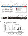
Imprinting status in the PWS/AS region (Chr. 15q11~13) and UBE3A expression. (A) Schematic representation of the PWS and AS region and primers designed to determine methylation imprints at the IC. PWS: Prader-Willi syndrome, AS: Angelman syndrome, IC: imprinting center. (B) Methylation-specific PCR analysis of the PWS-IC in all somatic and parthenogenetic cells. (C) Quantitative real-time PCR analysis of UBE3A expression in somatic and parthenogenetic cells during neural differentiation. The expression in hSiPSCs was set to 1. Means±SEM are shown for three independent experiments. Significance of inter-group differences was determined by one-way analysis of variance; ##p<0.01, ###p<0.001. Significance of the differences between cell types was determined by t-test, and p-values are indicated with *, **, and ***.

Similar articles

Cited by

References

    1. Reik W, Walter J. Genomic imprinting: parental influence on the genome. Nat Rev Genet. 2001;2:21–32. doi: 10.1038/35047554. - DOI - PubMed
    1. Plasschaert RN, Bartolomei MS. Genomic imprinting in development, growth, behavior and stem cells. Development. 2014;141:1805–1813. doi: 10.1242/dev.101428. - DOI - PMC - PubMed
    1. Battaglia A. The inv dup(15) or idic(15) syndrome: a clinically recognisable neurogenetic disorder. Brain Dev. 2005;27:365–369. doi: 10.1016/j.braindev.2004.08.006. - DOI - PubMed
    1. Woodfine K, Huddleston JE, Murrell A. Quantitative analysis of DNA methylation at all human imprinted regions reveals preservation of epigenetic stability in adult somatic tissue. Epigenetics Chromatin. 2011;4:1. doi: 10.1186/1756-8935-4-1. - DOI - PMC - PubMed
    1. Gregg C, Zhang J, Weissbourd B, Luo S, Schroth GP, Haig D, Dulac C. High-resolution analysis of parent-of-origin allelic expression in the mouse brain. Science. 2010;329:643–648. doi: 10.1126/science.1190830. - DOI - PMC - PubMed

LinkOut - more resources