

Classification of the human THAP protein family identifies an evolutionarily conserved coiled coil region
- PMID: 30836974
- PMCID: PMC6402169
- DOI: 10.1186/s12900-019-0102-2
Classification of the human THAP protein family identifies an evolutionarily conserved coiled coil region
Erratum in
-
Correction to: Classification of the human THAP protein family identifies an evolutionarily conserved coiled coil region.BMC Struct Biol. 2019 Mar 29;19(1):7. doi: 10.1186/s12900-019-0105-z. BMC Struct Biol. 2019. PMID: 30925869 Free PMC article.
Abstract
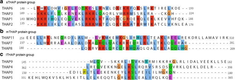
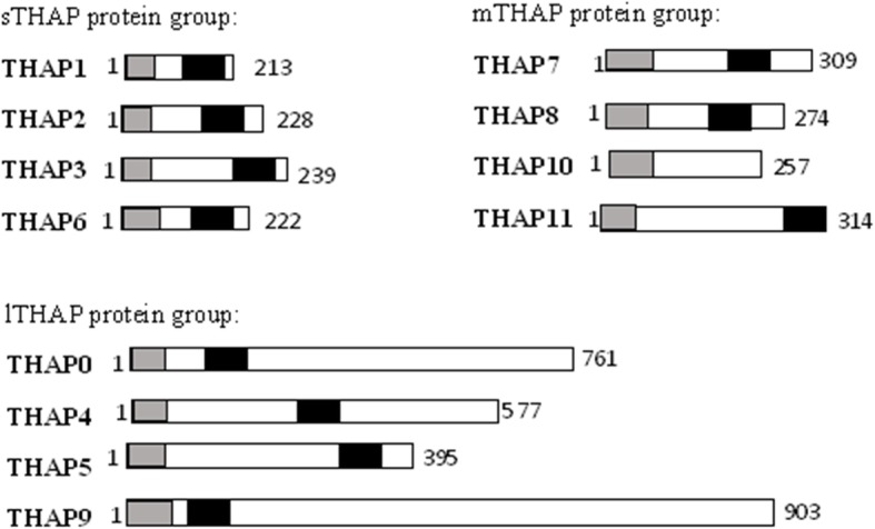
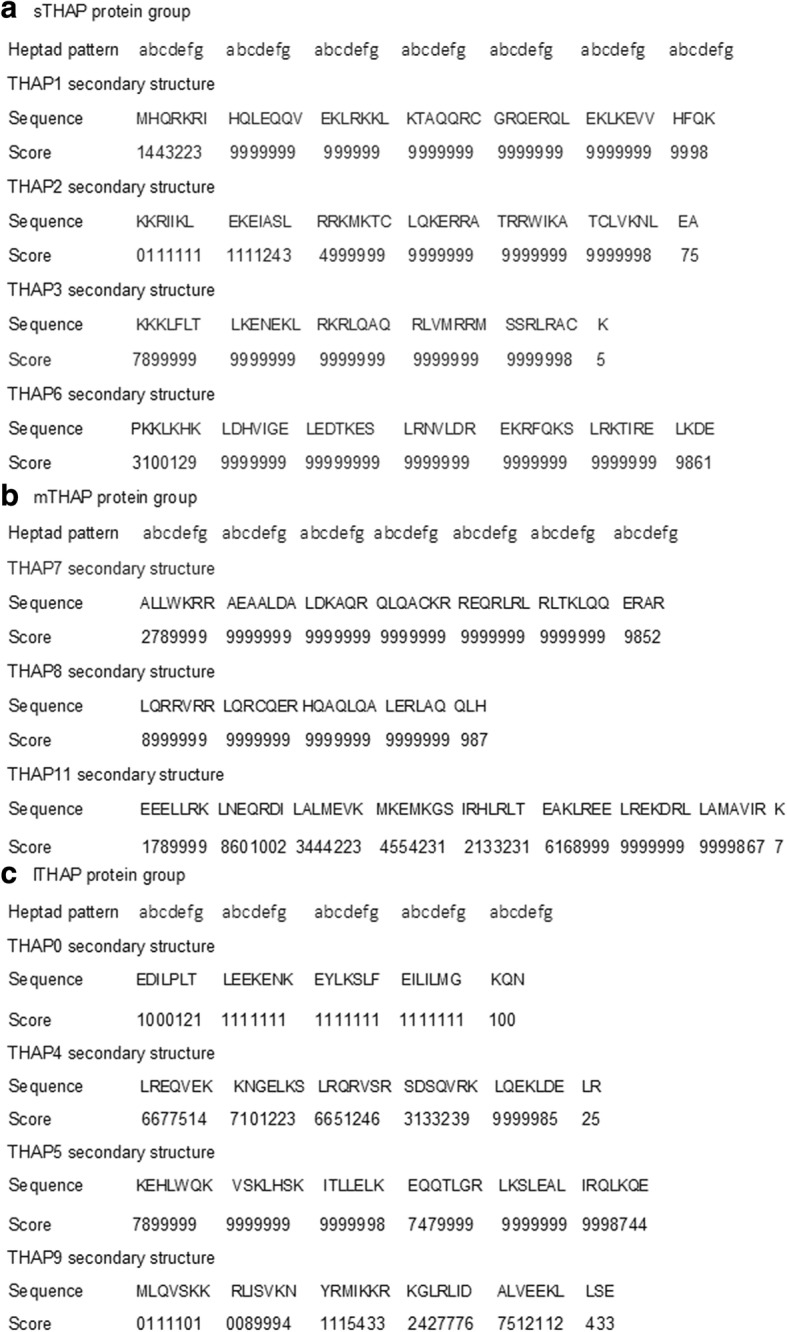
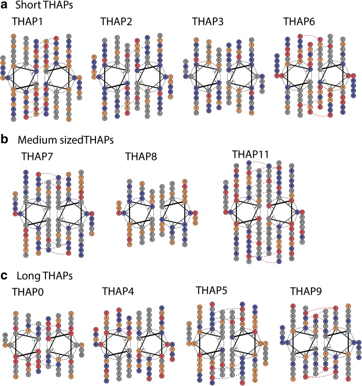
Background: The THAP (Thanatos Associated Proteins) protein family in humans is implicated in various important cellular processes like epigenetic regulation, maintenance of pluripotency, transposition and disorders like cancers and hemophilia. The human THAP protein family which consists of twelve members of different lengths has a well characterized amino terminal, zinc-coordinating, DNA-binding domain called the THAP domain. However, the carboxy terminus of most THAP proteins is yet to be structurally characterized. A coiled coil region is known to help in protein oligomerization in THAP1 and THAP11. It is not known if other human THAP proteins oligomerize. We have used bioinformatic tools to explore the possibility of dimerization of THAP proteins via a coiled coil region.
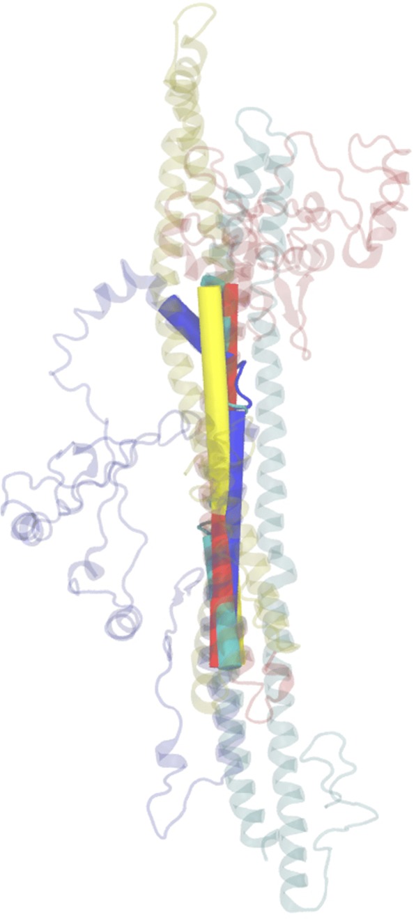
Results: Classification of human THAP protein into three size based groups led to the identification of an evolutionarily conserved alpha helical region, downstream of the amino terminal THAP domain. Secondary structure predictions, alpha helical wheel plots and protein models demonstrated the strong possibility of coiled coil formation in this conserved, leucine rich region of all THAP proteins except THAP10.
Conclusions: The identification of a predicted oligomerization region in the human THAP protein family opens new directions to investigate the members of this protein family.
Keywords: Classification; Leucine zipper; Oligomerization; THAP proteins.
Conflict of interest statement
Ethics approval and consent to participate
Not applicable
Consent for publication
Not applicable
Competing interests
The authors declare that they have no competing interests.
Publisher’s Note
Springer Nature remains neutral with regard to jurisdictional claims in published maps and institutional affiliations.
Figures

Similar articles
-
The C-terminal region of the transcriptional regulator THAP11 forms a parallel coiled-coil domain involved in protein dimerization.J Struct Biol. 2016 Jun;194(3):337-46. doi: 10.1016/j.jsb.2016.03.010. Epub 2016 Mar 11. J Struct Biol. 2016. PMID: 26975212
-
Dimerization of the DYT6 dystonia protein, THAP1, requires residues within the coiled-coil domain.J Neurochem. 2011 Sep;118(6):1087-100. doi: 10.1111/j.1471-4159.2011.07386.x. Epub 2011 Aug 8. J Neurochem. 2011. PMID: 21752024 Free PMC article.
-
In-depth Characterization of the Homodimerization Domain of the Transcription Factor THAP1 and Dystonia-Causing Mutations Therein.J Mol Neurosci. 2017 May;62(1):11-16. doi: 10.1007/s12031-017-0904-2. Epub 2017 Mar 15. J Mol Neurosci. 2017. PMID: 28299530
-
NMR studies of a new family of DNA binding proteins: the THAP proteins.J Biomol NMR. 2013 May;56(1):3-15. doi: 10.1007/s10858-012-9699-1. Epub 2013 Jan 11. J Biomol NMR. 2013. PMID: 23306615 Review.
-
Pharmacological interference with protein-protein interactions mediated by coiled-coil motifs.Handb Exp Pharmacol. 2008;(186):461-82. doi: 10.1007/978-3-540-72843-6_19. Handb Exp Pharmacol. 2008. PMID: 18491064 Review.
Cited by
-
THAP11 Functions as a Tumor Suppressor in Gastric Cancer through Regulating c-Myc Signaling Pathways.Biomed Res Int. 2020 Aug 27;2020:7838924. doi: 10.1155/2020/7838924. eCollection 2020. Biomed Res Int. 2020. PMID: 32908912 Free PMC article.
-
Correction to: Classification of the human THAP protein family identifies an evolutionarily conserved coiled coil region.BMC Struct Biol. 2019 Mar 29;19(1):7. doi: 10.1186/s12900-019-0105-z. BMC Struct Biol. 2019. PMID: 30925869 Free PMC article.
-
THAP11-mediated K48- and K63-linked ubiquitination is essential for the degradation of porcine reproductive and respiratory syndrome virus nonstructural protein 1β.Cell Mol Life Sci. 2025 Jun 23;82(1):246. doi: 10.1007/s00018-025-05760-3. Cell Mol Life Sci. 2025. PMID: 40548980 Free PMC article.
-
Bioinformatic analysis of THAP9 transposase homolog: conserved regions, novel motifs.Curr Res Struct Biol. 2023 Nov 24;7:100113. doi: 10.1016/j.crstbi.2023.100113. eCollection 2024. Curr Res Struct Biol. 2023. PMID: 38292821 Free PMC article.
-
A pathogenic DYT-THAP1 dystonia mutation causes hypomyelination and loss of YY1 binding.Hum Mol Genet. 2022 Mar 31;31(7):1096-1104. doi: 10.1093/hmg/ddab310. Hum Mol Genet. 2022. PMID: 34686877 Free PMC article.
References
-
- Macfarlan T, Kutney S, Altman B, Montross R, Yu J, Chakravarti D. Human THAP7 Is a Chromatin-associated, Histone Tail-binding Protein That Represses Transcription via Recruitment of HDAC3 and Nuclear Hormone Receptor Corepressor. J Biol Chem. 2004;280:7346–7358. doi: 10.1074/jbc.M411675200. - DOI - PubMed
-
- Cayrol C, Lacroix C, Mathe C, Ecochard V, Ceribelli M, Loreau E, Lazar V, Dessen P, Mantovani R, Aguilar L, Girard J-P. The THAP-zinc finger protein THAP1 regulates endothelial cell proliferation through modulation of pRB/E2F cell-cycle target genes. Blood. 2007;109:584–594. doi: 10.1182/blood-2006-03-012013. - DOI - PubMed
Publication types
MeSH terms
Substances
LinkOut - more resources
Full Text Sources
