Case Reports
. 2019 Mar 4;12(3):e228032.
doi: 10.1136/bcr-2018-228032.

Large air-filled intrapulmonary bronchogenic cyst associated with tension pneumothorax during air travel

Affiliations
Case Reports

Large air-filled intrapulmonary bronchogenic cyst associated with tension pneumothorax during air travel

Nicholas Bayfield et al. BMJ Case Rep. .

Abstract

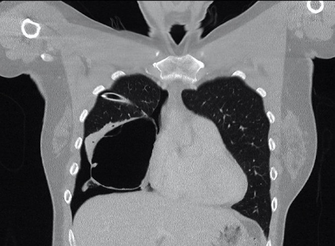
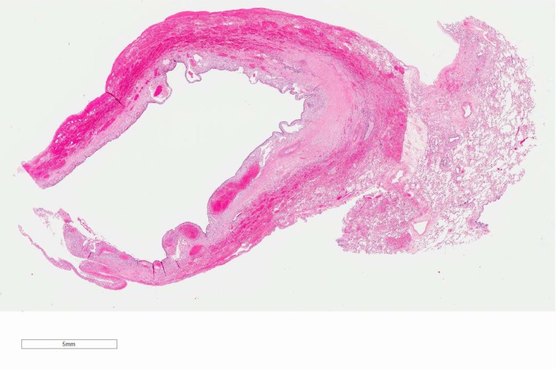
A 38-year-old woman developed a spontaneous right-sided tension pneumothorax during light aircraft travel. The aircraft was diverted to a regional centre, where emergent needle thoracostomy and chest tube insertion were performed. History suggested that this was the second episode of pneumothorax, with an untreated event with similar symptomatology during air travel 1 year ago. She was taken for surgical intervention. Intraoperative findings were of a large right middle lobe cyst of uncertain origin; the procedure was subsequently aborted. A CT chest demonstrated a large multiseptated air-filled pulmonary cystic lesion. Inpatient stay was notable for persistent right pneumothorax with interval cyst rupture. A right middle lobectomy was subsequently performed with histopathology showing a benign epithelioid bronchogenic cyst. Recovery was unremarkable with no residual pneumothorax or further episodes at 2 months postoperatively. Preventative excision of air-filled pulmonary abnormalities should be considered prior to air travel.

Keywords: air leaks; cardiothoracic surgery; pneumothorax; respiratory medicine.


Conflict of interest statement

Competing interests: None declared.

Figures

Figure 1
Figure 1
Chest X-ray demonstrating right-sided pneumothorax with leftwards mediastinal shift consistent with tension pneumothorax.
Figure 2
Figure 2
Interval right-sided chest drain insertion. Persistent right-sided pneumothorax with the resolution of tension features.
Figure 3
Figure 3
Chest X-ray reported as showing fluid in the oblique fissure, demonstrating a cystic structure in the right lower zone and small right-sided pneumothorax with chest drain in situ.
Figure 4
Figure 4
Axial CT slice showing an air-filled intrapulmonary right middle lobar cystic lesion with adjacent atelectasis and right-sided pneumothorax with chest drain in situ.
Figure 5
Figure 5
Coronal CT slice showing a large multiseptated air-filled intrapulmonary cystic lesion with adjacent atelectasis and right-sided pneumothorax with chest drain in situ.
Figure 6
Figure 6
Chest X-ray demonstrating interval expansion of right-sided pneumothorax with collapse of the cystic structure. Chest drain remains in situ.
Figure 7
Figure 7
Macroscopic pathological sectioning. Specimen type: right middle lobe. Specimen dimensions: a wedge of lung 105×50×45 mm. Number of lesions: one, unilocular collapsed cyst. Macroscopic cyst dimensions: 90×80×60 mm. Description of overlying pleura: congested and cream appearance. Non-neoplastic lung: unremarkable.
Figure 8
Figure 8
Microscopic (10× zoom) slide of the right middle lobe cyst. Sections showing lung parenchyma with a unilocular cyst lined by benign respiratory epithelium. The subepithelial stroma is fibrotic with focal condensation of bland myofibroblastic cells. There is no primitive or mesenchymal tissue associated with the cyst. There is evidence of old haemorrhage with some haemosiderin-laden macrophages. There is also blood clot material with a few mixed inflammatory cells including a few foreign body type giant cells and haemosiderin-laden macrophages. The background lung parenchyma appears unremarkable with no other lesions identified. Conclusion: benign respiratory epithelial lined cyst favoured to represent a bronchogenic cyst. There are no features to suggest malignancy.
Figure 9
Figure 9
Microscopic (200× zoom) slide of the right middle lobe cyst. Sections showing lung parenchyma with a unilocular cyst lined by benign respiratory epithelium. The subepithelial stroma is fibrotic with focal condensation of bland myofibroblastic cells. There is no primitive or mesenchymal tissue associated with the cyst. There is evidence of old haemorrhage with some haemosiderin-laden macrophages. There is also blood clot material with a few mixed inflammatory cells including a few foreign body type giant cells and haemosiderin-laden macrophages. The background lung parenchyma appears unremarkable with no other lesions identified. Conclusion: benign respiratory epithelial lined cyst favoured to represent a bronchogenic cyst. There are no features to suggest malignancy.
Figure 10
Figure 10
Microscopic (200× zoom) slide of the right middle lobe cyst. Sections showing lung parenchyma with a unilocular cyst lined by benign respiratory epithelium. The subepithelial stroma is fibrotic with focal condensation of bland myofibroblastic cells. There is no primitive or mesenchymal tissue associated with the cyst. There is evidence of old haemorrhage with some haemosiderin-laden macrophages. There is also blood clot material with a few mixed inflammatory cells including a few foreign body type giant cells and haemosiderin-laden macrophages. The background lung parenchyma appears unremarkable with no other lesions identified. Conclusion: benign respiratory epithelial lined cyst favoured to represent a bronchogenic cyst. There are no features to suggest malignancy.
Figure 11
Figure 11
Microscopic (400× zoom) slide of the right middle lobe cyst. Sections showing lung parenchyma with a unilocular cyst lined by benign respiratory epithelium. The subepithelial stroma is fibrotic with focal condensation of bland myofibroblastic cells. There is no primitive or mesenchymal tissue associated with the cyst. There is evidence of old haemorrhage with some haemosiderin-laden macrophages. There is also blood clot material with a few mixed inflammatory cells including a few foreign body type giant cells and haemosiderin-laden macrophages. The background lung parenchyma appears unremarkable with no other lesions identified. Conclusion: benign respiratory epithelial lined cyst favoured to represent a bronchogenic cyst. There are no features to suggest malignancy.

References

    1. Parkin L, Bell ML, Herbison GP, et al. . Air travel and fatal pulmonary embolism. Thromb Haemost 2006;95:807–14. - PubMed
    1. Pérez-Rodríguez E, Jiménez D, Díaz G, et al. . Incidence of air travel-related pulmonary embolism at the Madrid-Barajas airport. Arch Intern Med 2003;163:2766–70. 10.1001/archinte.163.22.2766 - DOI - PubMed
    1. Lapostolle F, Surget V, Borron SW, et al. . Severe pulmonary embolism associated with air travel. N Engl J Med 2001;345:779–83. 10.1056/NEJMoa010378 - DOI - PubMed
    1. Hu X, Cowl CT, Baqir M, et al. . Air travel and pneumothorax. Chest 2014;145:688–94. 10.1378/chest.13-2363 - DOI - PubMed
    1. Beunderman R, Sramek M, Koster RW, et al. . [Criteria for differential diagnosis in cardial symptoms; left- or right-sided chest pain?]. Ned Tijdschr Geneeskd 1990;134:2249–52. - PubMed

Publication types

MeSH terms