. 2019 Apr 26;294(17):6796-6808.
doi: 10.1074/jbc.RA118.006917. Epub 2019 Mar 5.

Global biochemical and structural analysis of the type IV pilus from the Gram-positive bacterium Streptococcus sanguinis

Affiliations

Global biochemical and structural analysis of the type IV pilus from the Gram-positive bacterium Streptococcus sanguinis

Jamie-Lee Berry et al. J Biol Chem. .

Abstract

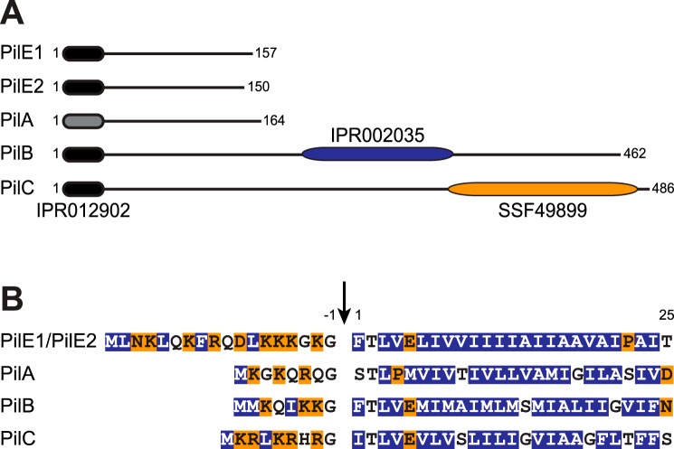
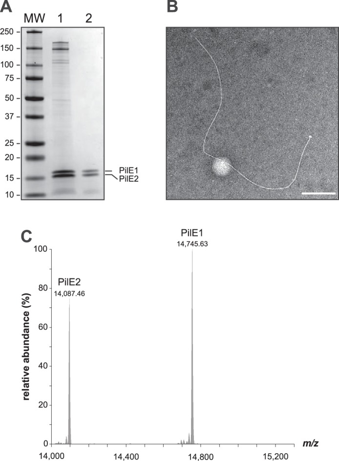
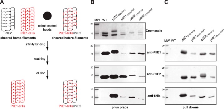
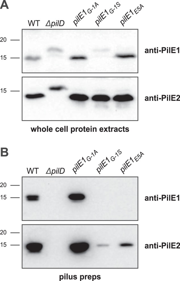
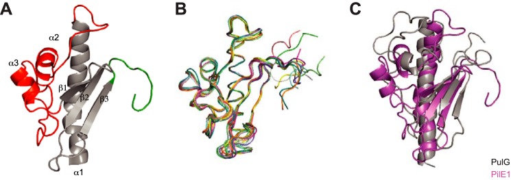
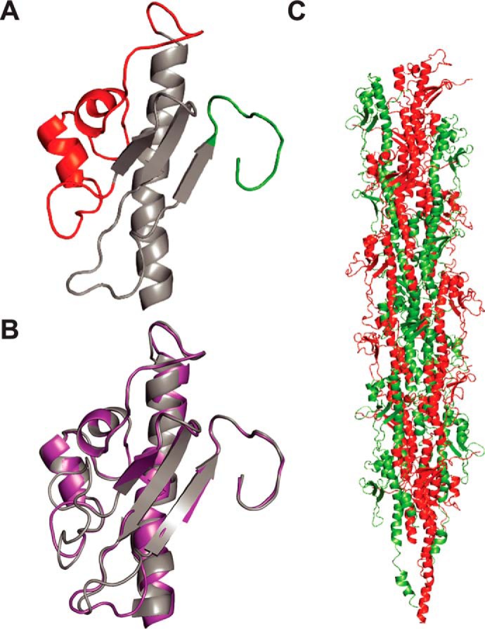
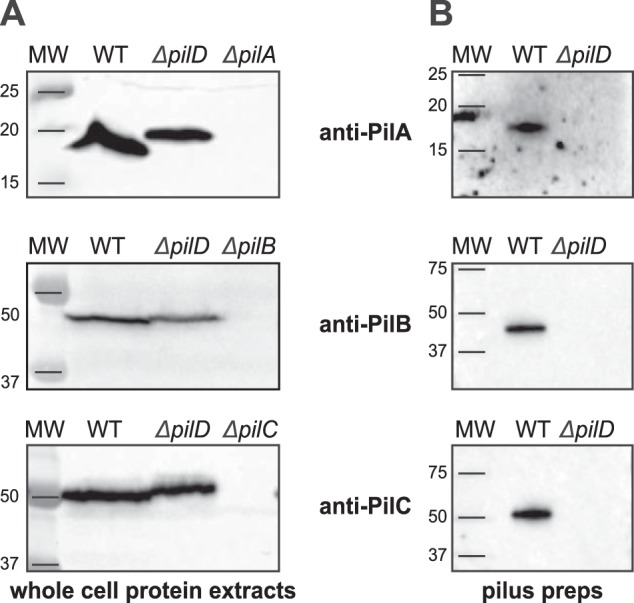
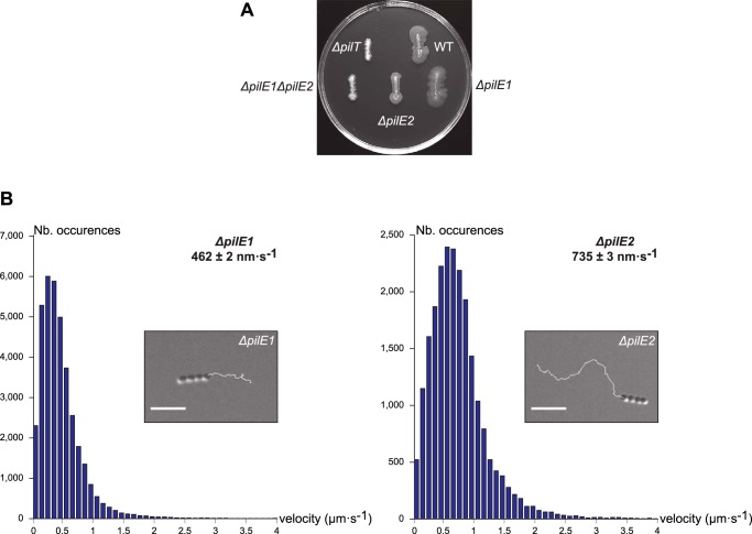
Type IV pili (Tfp) are functionally versatile filaments, widespread in prokaryotes, that belong to a large class of filamentous nanomachines known as type IV filaments (Tff). Although Tfp have been extensively studied in several Gram-negative pathogens where they function as key virulence factors, many aspects of their biology remain poorly understood. Here, we performed a global biochemical and structural analysis of Tfp in a recently emerged Gram-positive model, Streptococcus sanguinis In particular, we focused on the five pilins and pilin-like proteins involved in Tfp biology in S. sanguinis We found that the two major pilins, PilE1 and PilE2, (i) follow widely conserved principles for processing by the prepilin peptidase PilD and for assembly into filaments; (ii) display only one of the post-translational modifications frequently found in pilins, i.e. a methylated N terminus; (iii) are found in the same heteropolymeric filaments; and (iv) are not functionally equivalent. The 3D structure of PilE1, solved by NMR, revealed a classical pilin-fold with a highly unusual flexible C terminus. Intriguingly, PilE1 more closely resembles pseudopilins forming shorter Tff than bona fide Tfp-forming major pilins, underlining the evolutionary relatedness among different Tff. Finally, we show that S. sanguinis Tfp contain a low abundance of three additional proteins processed by PilD, the minor pilins PilA, PilB, and PilC. These findings provide the first global biochemical and structural picture of a Gram-positive Tfp and have fundamental implications for our understanding of a widespread class of filamentous nanomachines.

Keywords: Gram-positive bacteria; Streptococcus sanguinis; molecular motor; pilin; protein structure; twitching motility; type IV filaments; type IV pili; virulence factor.


Conflict of interest statement

The authors declare that they have no conflicts of interest with the contents of this article

Figures

Figure 1.
Figure 1.
Bioinformatic analysis of the two major pilins (PilE1 and PilE2) and three pilin-like proteins (PilA, PilB, and PilC) encoded by the pil locus in S. sanguinis 2908. A, protein architecture of the Pil proteins harboring the N-terminal IPR012902 motif that is part of the class III signal peptide defining type IV pilins (black rounded rectangle). In PilA, this motif could be detected only by visual inspection and is therefore represented by a gray rounded rectangle. PilB and PilC contain additional C-terminal domains (blue and orange rounded rectangles). PilB contains a von Willebrand factor type A motif (IPR002035), whereas PilC contains a concanavalin A-like lectin/glucanase structural domain (SSF49899). Proteins have been drawn to scale and the subscript numbers indicate their lengths. B, N-terminal sequence alignment of the putative class III signal peptides of the above five Pil proteins. The 8–18–aa long leader peptides, which contain a majority of hydrophilic (shaded in orange) or neutral (no shading) residues, end with a conserved Gly−1. Processing by PilD (indicated by the vertical arrow) is expected to occur after Gly−1. The mature proteins start with a tract of 21 predominantly hydrophobic residues (shaded in blue), which forms an extended α-helix that is the main assembly interface within filaments.
Figure 2.
Figure 2.
S. sanguinis Tfp exhibit a characteristic Tfp morphology and are composed primarily of N terminally methylated PilE1 and PilE2 subunits. A, SDS-PAGE/Coomassie analysis of S. sanguinis Tfp purified using previously described (lane 1) and enhanced (lane 2) purification procedures. Samples were prepared from cultures at similar OD600, separated by SDS-PAGE and stained with Coomassie Blue. Identical volumes were loaded in each lane. MW indicates the molecular mass marker lane, with masses in kDa. B, filament morphology in WT pilus preparations prepared using the enhanced purification procedure as assessed by TEM after negative staining. Scale bar represents 200 nm. C, top-down mass profiling of purified Tfp from S. sanguinis. Shown are deconvoluted molecular mass spectra over the range of 14,000 to 15,300 m/z. Masses are presented as monoisotopic [M + H]+.
Figure 3.
Figure 3.
S. sanguinis Tfp are heteropolymers composed of PilE1 and PilE2. A, schematics of the affinity-purification strategy. In brief, one of the genes encoding major pilin PilE1 (red) or PilE2 (black) is engineered to produce a protein fused to an affinity His6 tag, e.g. PilE1–6His (small red sphere). Sheared pili are mixed with cobalt-coated beads (large black sphere) and purified by pulldown, which is expected to yield filaments containing both pilins (tagged and untagged) if S. sanguinis Tfp are heteropolymers. Conversely, if the filaments are distinct homopolymers, only the tagged filaments will be purified. B, analysis of filaments purified by shearing/ultracentrifugation from four unmarked mutants harboring a His6 tag fused either to the C terminus of full-length PilE1 and PilE2 (PilE16His-long and PilE26His-long) or replacing the last seven aa in these pilins (PilE16His-short and PilE26His-short). The WT strain was included as a control. Samples were prepared from cultures at similar OD600, separated by SDS-PAGE and either stained with Coomassie Blue (upper panel) or analyzed by immunoblot using anti-PiE1, anti-PilE2, or anti-His6 antibodies (bottom three panels). MW indicates the molecular mass marker lane, in kDa. C, immunoblot analysis using anti-PiE1, anti-PilE2, or anti-His6 antibodies of sheared filaments that were affinity purified by pulldown. The WT strain was included as a control. Sheared filaments were prepared from cultures adjusted to the same OD600, affinity purified, eluted in the same final volume, and identical volumes were loaded in each lane.
Figure 4.
Figure 4.
Processing of S. sanguinis major pilin PilE1 by PilD and its assembly in filaments follow widely conserved general principles. A, immunoblot analysis of PilE1 expression and processing by PilD in strains expressing PilE1G−1A, PilE1G−1S, and PilE1E5A mutants. The WT strain and ΔpilD mutant have been included as controls. Whole cell protein extracts were separated by SDS-PAGE and probed using anti-PilE1 antibody or anti-PilE2 antibody as a control. Protein extracts were quantified, equalized, and identical volumes were loaded in each lane. Molecular masses are indicated in kDa. B, immunoblot analysis of PilE1 assembly in filaments in strains expressing PilE1G−1A, PilE1G−1S, and PilE1E5A mutants. The WT strain and ΔpilD mutant have been included as controls. Purified Tfp were separated by SDS-PAGE and probed using anti-PilE1 antibody or anti-PilE2 antibody as a control. Samples were prepared from cultures adjusted to the same OD600, and identical volumes were loaded in each lane but to improve detection of faint bands, we loaded 20-fold dilutions of WT and PilE1G−1A samples.
Figure 5.
Figure 5.
High-resolution 3D structure of PilE1 reveals a canonical type IV pilin-fold with an uncommon highly flexible C terminus, which more closely resembles pseudopilins. A, cartoon representation of the NMR structure of the globular domain of PilE1. The conserved core in type IV pilins (the N-terminal α-helix and 3-stranded antiparallel β-meander) is depicted in gray. Distinctive/key structural features flanking the β-meander are highlighted in color, the α1β1-loop in red, and the unstructured C terminus in green. B, ribbon representation of the superposition of the ensemble of 10 most favorable PilE1 structures determined by NMR. C, superimposed cartoon representations of the globular domains of PilE1 (magenta) and K. oxytoca pseudopilin PulG (gray). The two structures superpose with a root mean square deviation of 5.75 Å over their entire length.
Figure 6.
Figure 6.
Modeling shows that full-length PilE1/PilE2 fit readily into available Tfp structures. A, cartoon representation of the homology structural model of PilE2 based on PilE1 structure. The same color codes have been used as described in the legend to Fig. 5A. B, superposition of globular domains of PilE1 and PilE2 (right) with a root mean square deviation of 0.61 Å. C, packing of full-length models of PilE1 and PilE2 into the recently determined structure of gonoccoccal Tfp. Half of the gonoccoccal subunits in the structure were replaced by PilE1 and the other half with PilE2.
Figure 7.
Figure 7.
Pilin-like proteins PilA, PilB, and PilC are processed by PilD and co-purify with Tfp. A, immunoblot analysis of PiA, PilB, and PilC expression and processing by PilD. Whole-cell protein extracts were probed using specific antibodies, which were generated for this study. Protein extracts were quantified, equalized, and equivalent amounts of total proteins were loaded in each lane. Molecular masses are indicated in kDa. B, immunoblot analysis of pilus preparations using anti-PiA, anti-PilB, and anti-PilC antibodies. Samples were prepared from cultures adjusted to the same OD600 and identical volumes were loaded in each lane.
Figure 8.
Figure 8.
ΔpilE1 and ΔpilE2 mutants produce Tfp capable of powering motility, albeit at different speeds. A, macroscopic motility assay. Spreading zones, or lack thereof, around single and double ΔpilE1 and ΔpilE2 mutants. The WT strain and ΔpilT mutant have been included as positive and negative controls, respectively. B, microscopic motility assay. The histograms represents the distribution curve of velocities (in 100 nm s−1 intervals) measured for ΔpilE1 and ΔpilE2 mutants. Insets are representative 30-s trajectories of movement of small chains of cells. Scale bar represents 5 μm. Corresponding movies are available as Supplemental Information (Movies S1 and S2).

Similar articles

Cited by

References

    1. Berry J. L., and Pelicic V. (2015) Exceptionally widespread nano-machines composed of type IV pilins: the prokaryotic Swiss Army knives. FEMS Microbiol. Rev. 39, 134–154 10.1093/femsre/fuu001 - DOI - PMC - PubMed
    1. Chaudhury P., Quax T. E. F., and Albers S. V. (2018) Versatile cell surface structures of archaea. Mol. Microbiol. 107, 298–311 10.1111/mmi.13889 - DOI - PubMed
    1. Giltner C. L., Nguyen Y., and Burrows L. L. (2012) Type IV pilin proteins: versatile molecular modules. Microbiol. Mol. Biol. Rev. 76, 740–772 10.1128/MMBR.00035-12 - DOI - PMC - PubMed
    1. Pelicic V. (2008) Type IV pili: e pluribus unum? Mol. Microbiol. 68, 827–837 10.1111/j.1365-2958.2008.06197.x - DOI - PubMed
    1. Francetic O., Buddelmeijer N., Lewenza S., Kumamoto C. A., and Pugsley A. P. (2007) Signal recognition particle-dependent inner membrane targeting of the PulG pseudopilin component of a type II secretion system. J. Bacteriol. 189, 1783–1793 10.1128/JB.01230-06 - DOI - PMC - PubMed

Publication types