. 2019 Mar 5;10(2):e02807-18.
doi: 10.1128/mBio.02807-18.

Distinct Physiological Roles of the Three Ferredoxins Encoded in the Hyperthermophilic Archaeon Thermococcus kodakarensis

Affiliations

Distinct Physiological Roles of the Three Ferredoxins Encoded in the Hyperthermophilic Archaeon Thermococcus kodakarensis

Brett W Burkhart et al. mBio. .

Abstract

Control of electron flux is critical in both natural and bioengineered systems to maximize energy gains. Both small molecules and proteins shuttle high-energy, low-potential electrons liberated during catabolism through diverse metabolic landscapes. Ferredoxin (Fd) proteins-an abundant class of Fe-S-containing small proteins-are essential in many species for energy conservation and ATP production strategies. It remains difficult to model electron flow through complicated metabolisms and in systems in which multiple Fd proteins are present. The overlap of activity and/or limitations of electron flux through each Fd can limit physiology and metabolic engineering strategies. Here we establish the interplay, reactivity, and physiological role(s) of the three ferredoxin proteins in the model hyperthermophile Thermococcus kodakarensis We demonstrate that the three loci encoding known Fds are subject to distinct regulatory mechanisms and that specific Fds are utilized to shuttle electrons to separate respiratory and energy production complexes during different physiological states. The results obtained argue that unique physiological roles have been established for each Fd and that continued use of T. kodakarensis and related hydrogen-evolving species as bioengineering platforms must account for the distinct Fd partnerships that limit flux to desired electron acceptors. Extrapolating our results more broadly, the retention of multiple Fd isoforms in most species argues that specialized Fd partnerships are likely to influence electron flux throughout biology.IMPORTANCE High-energy electrons liberated during catabolic processes can be exploited for energy-conserving mechanisms. Maximal energy gains demand these valuable electrons be accurately shuttled from electron donor to appropriate electron acceptor. Proteinaceous electron carriers such as ferredoxins offer opportunities to exploit specific ferredoxin partnerships to ensure that electron flux to critical physiological pathways is aligned with maximal energy gains. Most species encode many ferredoxin isoforms, but very little is known about the role of individual ferredoxins in most systems. Our results detail that ferredoxin isoforms make largely unique and distinct protein interactions in vivo and that flux through one ferredoxin often cannot be recovered by flux through a different ferredoxin isoform. The results obtained more broadly suggest that ferredoxin isoforms throughout biological life have evolved not as generic electron shuttles, but rather serve as selective couriers of valuable low-potential electrons from select electron donors to desirable electron acceptors.

Keywords: Thermococcus kodakarensis; archaea; electron flux; ferredoxin; hydrogenase; hyperthermophile.


Figures

FIG 1
FIG 1
T. kodakarensis encodes three ferredoxins with dramatically different physical properties and expression patterns. (A) Sequence alignments of Fd-1, Fd-2, and Fd-3, highlighting residues likely involved in metal coordination (blue). (B, C, and D) Cartoon Phyre2 models of Fd-1 (B), Fd-2 (C), and Fd-3 (D) with electrostatic surface potentials shown below from two perspectives. (E) Summary of physical properties, essentiality, and genome organization of each Fd.
FIG 2
FIG 2
Loci encoding each Fd can be modified within T. kodakarensis to encode proteins with C-terminal extensions without compromising viability or growth rate. (A) Map of the TK1694 locus in the genomes of TS559 (top) and TK1694-CT (bottom), highlighting the approximate location of primer binding sites used to generate amplicons that were resolved either prior to (−) or following digestion with BspEI. Lane M in panels A, B, and C contains size markers in kbp. C-plasmid DNA for TK1694 was used as a control to confirm BspEI digestion of amplicon DNA. (B) Map of the TK1087 locus in the genomes of TS559 (top) and TK1087-CT (bottom), highlighting the approximate location of primer binding sites used to generate amplicons that were resolved in the gel below. Primers 1 and 2 are complementary to flanking sequences, while primers 3 and 4 are complementary to sequences within the 45-bp tag sequence at the C terminus of TK1087. Amplification with primer pairs 1 and 2 generates near-equivalent-size amplicons from genomic DNA from strains TS559 and TK1087-CT, while only DNA from TK1087-CT supports generation of amplicons with primer pairs 3 and 2 or 1 and 4. (C) Map of the TK2012 locus in the genomes of TS559 (top) and TK2012-CT (bottom), highlighting the approximate location of primer binding sites used to generate amplicons that were resolved either prior to (−) or following digestion with BspEI. C-plasmid DNA for TK2012 was used as a control to confirm BspEI digestion of amplicon DNA. (D) Strains with C-terminal extensions at individual Fd loci grow nearly identically to strain TS559 under standard laboratory conditions. The values plotted are the average of three assays each of triplicate cultures of each strain.
FIG 3
FIG 3
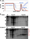
Fd proteins maintain stable interactions permitting copurification of Fd-binding partners. (A) Elution profiles of total clarified cell lysates of strains TS559 (black) and TK1694-CT (red) reveal an additional peak of absorbance at 280 nm (peak 2) that elutes at higher imidazole concentrations from lysates of TK1694-CT and which is not present in the parental strain, TS559. (B) SDS-PAGE of aliquots of fractions recovered from Ni2+-charged chelating chromatography from strains TS559 (top) and TK1694-CT (bottom) reveal near-identical protein patterns within peak 1, representing native T. kodakarensis proteins with mild affinity for the Ni2+-charged chelating matrix. Resolution of aliquots from peak 2 reveals minimal proteins in lysates derived from strain TS559, but obvious retention of Fd-1 (red arrow) and Fd-1-interacting proteins (red bracket) from lysates derived from strain TS1694-CT.
FIG 4
FIG 4
The T. kodakarensis Fd interactomes are distinct. The primary electron donors that reduced Fds are shown on the left within a model of glycolysis and amino acid fermentation. The primary electron acceptors that oxidize reduced Fds are shown to the right within partial representations of soluble lipid and NAD(P)H production pathways and membrane-bound respiratory and ATP-generating complexes. Each Fd is highlighted in the center of the panel, with interacting partners connected by solid, colored lines (Fd-1, green; Fd-2, pink; Fd-3, blue).
FIG 5
FIG 5
TK2012, encoding Fd-3, is nonessential in the presence of S0. (A) Map of the TK2012 locus in the genome of TS559 highlighting the approximate location of primer binding sites used to generate amplicons that were resolved in the gel below. Primers 1 and 2 are complementary to TK2012 flanking sequences, while primer 3 is complementary to sequences within TK2012. Amplification with primer pairs 1 and 2 generates a shorter amplicon from genomic DNA from the ΔTK2012 strain than DNA from strain TS559, reflecting the loss of TK2012 coding sequences. Only DNA from TS559 supports generation of an amplicon with primer pair 1 and 3. Lane M contains size markers in kbp. (B) Growth of strains TS559 and ΔTK2012 in the presence of S0 (lower panel) is robust, whereas the ΔTK2012 strain fails to achieve significant densities in the absence of S0 (top panel).

References

    1. Mayer F, Müller V. 2014. Adaptations of anaerobic archaea to life under extreme energy limitation. FEMS Microbiol Rev 38:449–472. doi:10.1111/1574-6976.12043. - DOI - PubMed
    1. Martin WF, Thauer RK. 2017. Energy in ancient metabolism. Cell 168:953–955. doi:10.1016/j.cell.2017.02.032. - DOI - PubMed
    1. Dillon JG, Anantharaman K, Lee Moyer C, Xiao X, Ding J, Zhang Y, Wang H, Jian H, Leng H. 2017. Microbial community structure of deep-sea hydrothermal vents on the ultraslow spreading Southwest Indian Ridge. Arctic Front Microbiol 8:1012. doi:10.3389/fmicb.2017.01012. - DOI - PMC - PubMed
    1. Adams MW. 1994. Biochemical diversity among sulfur-dependent, hyperthermophilic microorganisms. FEMS Microbiol Rev 15:261–277. doi:10.1111/j.1574-6976.1994.tb00139.x. - DOI - PubMed
    1. Schut GJ, Zadvornyy O, Wu C-H, Peters JW, Boyd ES, Adams MWW. 2016. The role of geochemistry and energetics in the evolution of modern respiratory complexes from a proton-reducing ancestor. Biochim Biophys Acta 1857:958–970. doi:10.1016/j.bbabio.2016.01.010. - DOI - PubMed

Publication types

MeSH terms

LinkOut - more resources