. 2019 Mar 5;9(1):3594.
doi: 10.1038/s41598-019-40136-1.

Vegfa/vegfr2 signaling is necessary for zebrafish islet vessel development, but is dispensable for beta-cell and alpha-cell formation

Affiliations

Vegfa/vegfr2 signaling is necessary for zebrafish islet vessel development, but is dispensable for beta-cell and alpha-cell formation

Chiara M Toselli et al. Sci Rep. .

Abstract

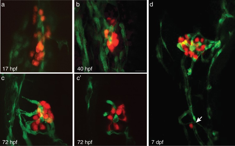
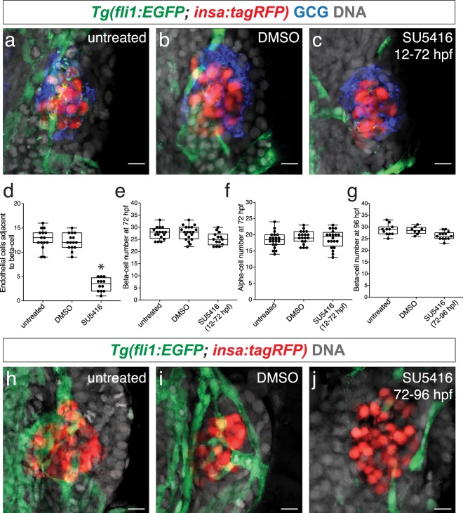
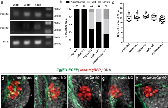
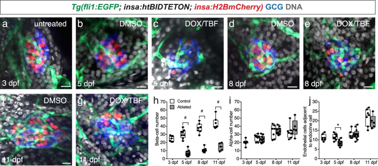
The mechanisms underlying zebrafish pancreatic islet vascularization have not been well characterized. We sought to determine the angiogenic factors responsible for islet vascularization and assess whether an absence of endothelial cells affects beta-cell and alpha-cell formation. We used a double transgenic zebrafish Tg(fli1:EGFP; insa:tagRFP) to label endothelial cells and beta-cells, respectively. Beta-cells developed adjacent to endothelial cells and by 72 hours post fertilization (hpf) the zebrafish pancreatic islet was highly vascularized. Zebrafish beta-cells express vascular endothelial growth factors (vegf), vegfaa and vegfab. Double knockdown of vegfaa and vegfab or the primary Vegfa receptors (Vegfr2), kdr and kdrl, resulted in vessel deficient islets. While beta-cell and alpha-cell numbers remained unchanged in vessel deficient islets, insulina expression was downregulated relative to controls. Vegfaa/Vegfab-Vegfr2 signaling is necessary for proper islet vessel development, but not for the initial formation of beta-cells and alpha-cells.


Conflict of interest statement

The authors declare no competing interests.

Figures

Figure 1
Figure 1
The endocrine pancreas develops adjacent to vessels and is highly vascularized. (a–c) Confocal projections of the pancreatic islet at 17 hpf, 40 hpf, and 72 hpf in Tg(fli1:EGFP; insa:tagRFP); endothelial cells (green) and beta-cells (red). (c’) Confocal section of projection in (c). (d) Confocal projection of 7 dpf Tg(fli1:EGFP; insa:tagRFP) pancreas. Arrow indicates secondary islet.
Figure 2
Figure 2
Inhibiting Vegf signaling does not affect beta-cell and alpha-cell formation. (a–c) Confocal projections of 72 hpf Tg(fli1:EGFP; insa:tagRFP) untreated, DMSO-treated, and SU5416-treated embryos from 12 to 72 hpf; endothelial cells (green), beta-cells (red), and DAPI nuclear stain (DNA; grey). Alpha-cells are labeled with a glucagon (GCG) antibody (blue). (d) The number of endothelial cells adjacent to beta-cells in untreated, DMSO-treated, and SU5416-treated embryos from 12 to 72 hpf. (e,f) The number of beta-cells and alpha-cells in Tg(fli1:EGFP; insa:tagRFP) untreated, DMSO-treated, and SU5416-treated embryos from 12 to 72 hpf. n = 14–20. (g) The number of beta-cells in Tg(fli1:EGFP; insa:tagRFP) untreated, DMSO-treated, and SU5416-treated embryos from 72 hpf to 92 hpf. n = 8–13. (h–j) Confocal projections of 96 hpf Tg(fli1:EGFP; insa:tagRFP) untreated, DMSO-treated, and SU5416-treated embryos from 72 to 96 hpf; endothelial cells (green), beta-cells (red), and DAPI (grey). (d–g) Box-and-whisker plots show median, and circles represent individual zebrafish. Scale bar = 10 μm.
Figure 3
Figure 3
Vegfaa and Vegfab are necessary for islet vessel development. (a) RT-PCR of vegfaa and vegfab on sorted 2 dpf, 3 dpf, and adult beta-cells. Full length gel is presented in Supplementary Fig. 3. (b) Phenotypic score of islet vessels in non-injected, control morpholino and vegfaa, vegfab, or vegfaa/vegfab morpholino injected Tg(fli1:EGFP; insa:tagRFP) embryos. Phenotypes are scored such that no phenotype is comparable to wildtype (more than 7 endothelial cells adjacent to beta-cells), mild phenotype (4–7 endothelial cells adjacent to beta-cells), and severe phenotype (less than 4 endothelial cells adjacent to beta-cells). (c) The number of beta-cells in 72 hpf Tg(fli1:EGFP; insa:tagRFP) control and morpholino injected embryos. In the vegfaa, vegfab, and vegfaa/ab morpholino injected embryos, only the embryos that demonstrated a reduction or absence of islet vessels were counted. n = 9–21. Box-and-whisker plots show median, and circles represent individual zebrafish. (d–h) Confocal projections of (d) non-injected, (e) scrambled injected, (f) vegfaa, (g) vegfab, or (h) vegfaa/ab morpholino injected embryos at 72 hpf; endothelial cells (green), beta-cells (red), and DAPI (grey). Scale bar = 10 μm.
Figure 4
Figure 4
Knockdown of Vegfr2 receptors kdr and kdrl leads to disruptions in islet vessel development. (a) Phenotypic score of islet vessels in non-injected, control morpholino, and kdr, kdrl, or kdr/kdrl morpholino injected Tg(fli1:EGFP; insa:tagRFP) embryos. (b) The number of beta-cells in 72 hpf Tg(fli1:EGFP; insa:tagRFP) control and morpholino injected embryos. In the kdr/kdrl morpholino injected embryos, only the embryos that demonstrated a reduction or absence of islet vessels were counted. Box-and-whisker plots show median, and circles represent individual zebrafish. n = 17–24. (c–e) Relative expression of (c) insa, (d) pdx1, and (e) neuroD in isolated beta-cells of control injected and kdr/kdrl morpholino injected Tg(fli1:EGFP; insa:tagRFP) embryos at 72 hpf. All values were normalized to ef1α. *p < 0.0001 by Student’s t-test. (f–j) Confocal projections of (f) non-injected, (g) control injected, (h) kdr, (i) kdrl, or (j) kdr/kdrl morpholino injected embryos at 72 hpf; endothelial cells (green), beta-cells (red), and DAPI (grey). Scale bar = 10 μm.
Figure 5
Figure 5
The number of islet endothelial cells decreases after beta-cell ablation. (a–g) Confocal projections of (a) 3 dpf, (b) 5 dpf control, (c) 5 dpf beta-cell ablated fish, (d) 8 dpf control, (e) 8 dpf beta-cell ablated fish, (f) 11 dpf control, (g) 11 dpf beta-cell ablated Tg(-1.2ins:htBidTE-ON; -1.2ins:H2BmCherry; fli1:EGFP) fish; endothelial cells (green), beta-cells (red), glucagon (blue) and DAPI (grey). Scale bar = 10 μm. (h–j) The number of (h) beta-cells, (i) alpha-cells, and (j) endothelial cells during beta-cell ablation and regeneration in Tg(-1.2ins:htBidTE-ON; -1.2ins:H2BmCherry; fli1:EGFP) fish treated with either DMSO (control) or DOX/TBF (beta-cell ablated) from 3–5 dpf. Box-and-whisker plots show median, and circles represent individual zebrafish. n = 5–12. Student’s t-test was conducted between the control and ablated groups at the same timepoint. #p < 0.0001, *p = 0.0004.

References

    1. Lammert E, Cleaver O, Melton D. Induction of Pancreatic Differentiation by Signals from Blood Vessles. Science. 2001;294:564–567. doi: 10.1126/science.1064344. - DOI - PubMed
    1. Yoshitomi H, Zaret KS. Endothelial cell interactions initiate dorsal pancreas development by selectively inducing the transcription factor Ptf1a. Development. 2004;131:807–817. doi: 10.1242/dev.00960. - DOI - PubMed
    1. Jacquemin P, et al. An endothelial-mesenchymal relay pathway regulates early phases of pancreas development. Dev. Biol. 2006;290:189–199. doi: 10.1016/j.ydbio.2005.11.023. - DOI - PubMed
    1. Brissova M, et al. Pancreatic islet production of vascular endothelial growth factor-A is essential for islet vascularization, revascularization, and function. Diabetes. 2006;55:2974–2985. doi: 10.2337/db06-0690. - DOI - PubMed
    1. Lammert E, et al. Role of VEGF-A in Vascularization of Pancreatic Islets. Curr. Biol. 2003;13:1070–1074. doi: 10.1016/S0960-9822(03)00378-6. - DOI - PubMed

Publication types

MeSH terms

Substances

LinkOut - more resources