. 2019 Mar 5;9(1):3533.
doi: 10.1038/s41598-019-40190-9.

Platelet lysate outperforms FCS and human serum for co-culture of primary human macrophages and hMSCs

Affiliations

Platelet lysate outperforms FCS and human serum for co-culture of primary human macrophages and hMSCs

Tina Tylek et al. Sci Rep. .

Abstract

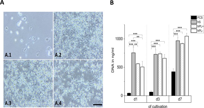
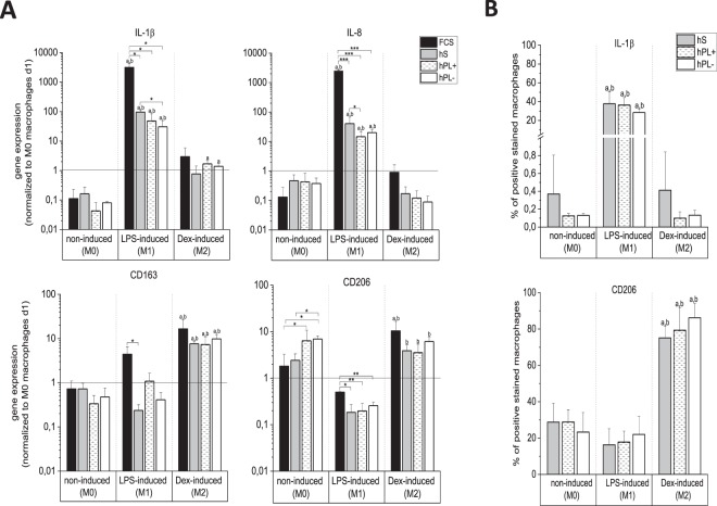
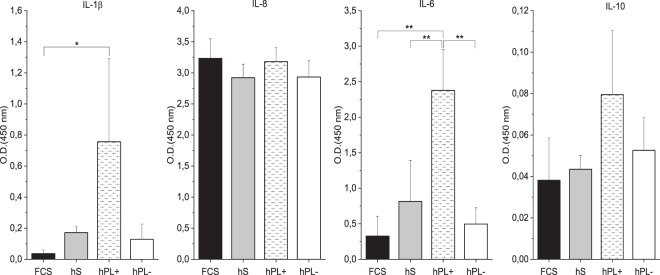
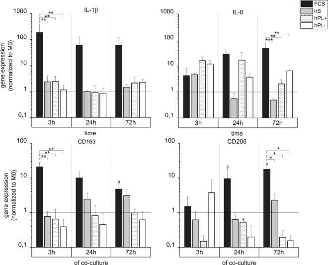
In vitro co-cultures of different primary human cell types are pivotal for the testing and evaluation of biomaterials under conditions that are closer to the human in vivo situation. Especially co-cultures of macrophages and mesenchymal stem cells (MSCs) are of interest, as they are both present and involved in tissue regeneration and inflammatory reactions and play crucial roles in the immediate inflammatory reactions and the onset of regenerative processes, thus reflecting the decisive early phase of biomaterial contact with the host. A co-culture system of these cell types might thus allow for the assessment of the biocompatibility of biomaterials. The establishment of such a co-culture is challenging due to the different in vitro cell culture conditions. For human macrophages, medium is usually supplemented with human serum (hS), whereas hMSC culture is mostly performed using fetal calf serum (FCS), and these conditions are disadvantageous for the respective other cell type. We demonstrate that human platelet lysate (hPL) can replace hS in macrophage cultivation and appears to be the best option for co-cultivation of human macrophages with hMSCs. In contrast to FCS and hS, hPL maintained the phenotype of both cell types, comparable to that of their respective standard culture serum, as well as the percentage of each cell population. Moreover, the expression profile and phagocytosis activity of macrophages was similar to hS.


Conflict of interest statement

The authors declare no competing interests.

Figures

Figure 1
Figure 1
Phenotype and adhesion of spontaneously differentiated macrophages. The phenotype (A) of macrophages after seven days of cultivation in medium with hS (A.2), hPL+ heparin (A.3), and hPL− heparin (A.4) was similar, while macrophages cultivated in medium with FCS showed a markedly different morphology (A.1). The analysis of DNA amounts (B) corresponding to cell numbers, provided information about cell adhesion behavior (mean ± SD, n = 3). Scale bar: 100 µm. **p < 0.01, ***p < 0.001.
Figure 2
Figure 2
Gene and protein expression of spontaneously (M0) and induced (M1/M2) differentiated macrophages after seven days. (A) Gene expression was analyzed by qPCR. Independent of culture conditions, spontaneous as well as induced differentiation was observed. The M1 markers IL-1β and IL-8 were downregulated in the non-induced (M0) as well as Dex-induced (M2) macrophages and highly upregulated in LPS-induced (M1) ones. The M2 markers CD163 and CD206 were more highly expressed in M2 macrophages, compared to M0 and M1. Spontaneous differentiation (M0) differed from the reference sample (monocytes/macrophages on day 1) in all four media (mean ± SD, n = 3). (B) Polarization into M0/M1/M2 macrophages determined via protein analysis using flow cytometry. M1 macrophages reflected a 40% higher IL-1β expression compared to M0 and M2. In contrast, M2 macrophages markedly showed a 60% higher protein expression of CD206 in comparison to M0 an M1 macrophages (mean ± SD, n = 3). *p < 0.05; **p < 0.01, ***p < 0.001; ap < 0.05 vs non-induced (M0), same serum supplement; bp < 0.05 between LPS-induced (M1)/Dex-induced (M2), same serum supplement.
Figure 3
Figure 3
Cytokine release profiles of spontaneously differentiated macrophages after seven days of culture. Cytokine release of IL-1β, IL-6, IL-8, and IL-10 was detected in supernatants of macrophages cultivated in medium with hS, FCS, and hPL+/− heparin using a commercially available ELSIA kit. Relative protein amounts are given by arbitrary units of optical density at 450 nm and show varying release profiles for different sera supplementations (mean ± SD, n = 3). *p < 0.05; **p < 0.01.
Figure 4
Figure 4
Phenotype and proportion of cell populations in the co-culture of M0 macrophages and hMSCs. The phenotypes of macrophages (green) and hMSCs (orange) were investigated under different culture conditions (A). In medium with FCS (A.1) only very few macrophages adhered, in medium with hS (A.2) hMSCs aggregated. In cell cultures with hPL+(A.3)/−(A.4) heparin, both, macrophages and hMSCs, displayed cell type-specific phenotypes corresponding to their morphology in single cultures with the respective standard serum. The percentage of macrophages in co-cultures measured via flow cytometric analysis of CD45 expression (B) confirmed the microscopic observations (mean ± SD, n = 4). Scale bar = 200 µm. ***p < 0.001.
Figure 5
Figure 5
Gene expression profile of M0 macrophages after co-cultivation with hMSCs. The gene expression of the M1 markers IL-1β and IL-8 as well as the M2 markers CD163 and CD206 was analyzed for CD45-positive macrophages after 3 h, 24 h and 72 h of co-cultivation by qPCR. For the different culture conditions (medium supplemented with hS/FCS/hPL+/−), expression levels are depicted with normalization to RPS27A and M0 macrophages (mean ± SD, n = 3). *p < 0.05; **p < 0.01; ***p < 0.001; ap < 0.05 vs 3 h, same serum supplement.
Figure 6
Figure 6
Phagocytic activity of M0 macrophages mono- and co-cultured with hMSCs. The phagocytic activity of CD45-positive macrophages under different culture conditions (medium with hS/FCS/hPL+ and − heparin) was measured via flow cytometry for the uptake of fluorescent beads after 3 h, 24 h, and 72 h. In medium with FCS and hPL, the phagocytosis rate of co-cultured cells exceeded that of the mono-cultured ones. Contrarily, in medium with hS, mono-cultured macrophages even exhibited a higher bead uptake than those in co-culture (mean ± SD, n = 3). **p < 0.01; ***p < 0.001.

References

    1. Gordon, S. & Taylor, P. R. Monocyte and macrophage heterogeneity. Nat Rev Immunol5, 953–964, 10.1038nri1733 (2005). - PubMed
    1. Shapouri-Moghaddam A, et al. Macrophage plasticity, polarization, and function in health and disease. J Cell Physiol. 2018;233:6425–6440. doi: 10.1002/jcp.26429. - DOI - PubMed
    1. Sica A, Mantovani A. Macrophage plasticity and polarization: in vivo veritas. J Clin Invest. 2012;122:787–795. doi: 10.1172/JCI59643. - DOI - PMC - PubMed
    1. Leopold Wager CM, Wormley FL., Jr. Classical versus alternative macrophage activation: the Ying and the Yang in host defense against pulmonary fungal infections. Mucosal Immunol. 2014;7:1023–1035. doi: 10.1038/mi.2014.65. - DOI - PubMed
    1. Martinez FO, Gordon S. The M1 and M2 paradigm of macrophage activation: time for reassessment. F1000Prime Rep. 2014;6:13. doi: 10.12703/P6-13. - DOI - PMC - PubMed

Publication types

MeSH terms