

B-cell depletion induces a shift in self antigen specific B-cell repertoire and cytokine pattern in patients with bullous pemphigoid
- PMID: 30837635
- PMCID: PMC6401188
- DOI: 10.1038/s41598-019-40203-7
B-cell depletion induces a shift in self antigen specific B-cell repertoire and cytokine pattern in patients with bullous pemphigoid
Erratum in
-
Author Correction: B-cell depletion induces a shift in self antigen specific B-cell repertoire and cytokine pattern in patients with bullous pemphigoid.Sci Rep. 2019 Dec 9;9(1):18991. doi: 10.1038/s41598-019-54421-6. Sci Rep. 2019. PMID: 31813939 Free PMC article.
Abstract
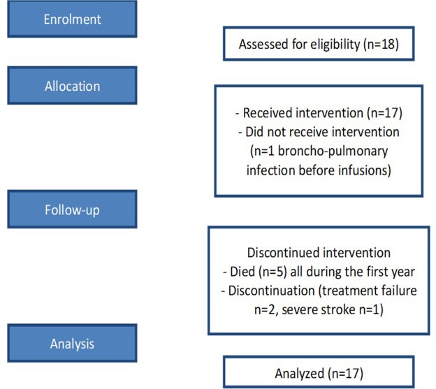
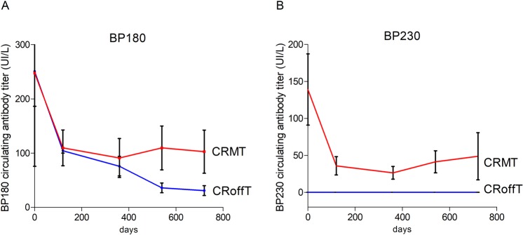
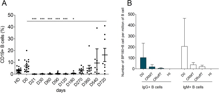
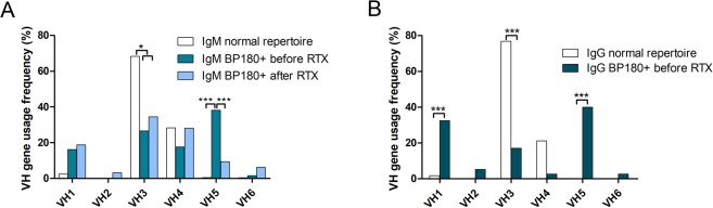
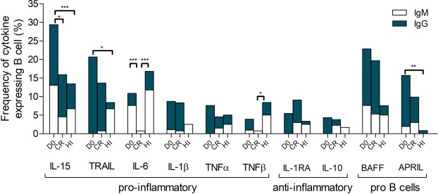
Bullous Pemphigoid is the most common auto-immune bullous skin disease. It is characterized by the production of auto-antibodies directed against 2 proteins of the hemi-desmosome (BP180 and BP230). We assessed the efficacy and mechanisms of action of rituximab, an anti-CD20 monoclonal antibody, in 17 patients with severe and relapsing type of bullous pemphigoid. The phenotype, cytokine gene expression, and rearrangement of BP180-specific B-cell receptor genes were performed over 2 years following treatment. At the end of the study, 5 patients had died, 3 had withdrawn from the study, and 9 patients were in complete remission. The one- and two-year relapse rates were 44.1% (95% Confidence Interval (CI): 21.0-76.0%) and 66.5%, (95% CI: 38.4-91.4%), respectively. Phenotypic analyses confirmed dramatic B-cell depletion, which lasted for 9 to 12 months. The ELISA values of serum anti-BP180 antibodies and the frequency of BP180-specific circulating B cells decreased dramatically following treatment, which paralleled the improvement of skin lesions. During B-cell reconstitution, a polyclonal IgM repertoire appeared and a shift in the rearrangement of the B-cell receptor genes of BP180-specific circulating B cells was observed. Concurrently, we observed a decrease of IL-15, IL-6 and TNFα expressing BP180-specific B cells, and the emergence of IL-10 and IL-1RA-expressing BP180-specific IgM+ B cells in patients in complete remission off therapy, suggesting the functional plasticity of BP180-specific auto-immune B cells after rituximab treatment.
Conflict of interest statement
The authors declare no competing interests.
Figures
Similar articles
-
Clinical and immunologic factors associated with bullous pemphigoid relapse during the first year of treatment: a multicenter, prospective study.JAMA Dermatol. 2014 Jan;150(1):25-33. doi: 10.1001/jamadermatol.2013.5757. JAMA Dermatol. 2014. PMID: 24226428
-
Autoreactive Peripheral Blood T Helper Cell Responses in Bullous Pemphigoid and Elderly Patients With Pruritic Disorders.Front Immunol. 2021 Mar 25;12:569287. doi: 10.3389/fimmu.2021.569287. eCollection 2021. Front Immunol. 2021. PMID: 33841390 Free PMC article.
-
Serum levels of IgE anti-BP180 and anti-BP230 autoantibodies in patients with bullous pemphigoid.J Dermatol Sci. 2008 Feb;49(2):153-61. doi: 10.1016/j.jdermsci.2007.08.008. Epub 2007 Oct 24. J Dermatol Sci. 2008. PMID: 17920818
-
IgE-mediated mechanisms in bullous pemphigoid and other autoimmune bullous diseases.Expert Rev Clin Immunol. 2016;12(3):267-77. doi: 10.1586/1744666X.2016.1123092. Epub 2015 Dec 16. Expert Rev Clin Immunol. 2016. PMID: 26588556 Review.
-
Immunoglobulin E and bullous pemphigoid.Eur J Dermatol. 2018 Aug 1;28(4):440-448. doi: 10.1684/ejd.2018.3366. Eur J Dermatol. 2018. PMID: 30325326 Review.
Cited by
-
Study of cytokine-induced immunity in bullous pemphigoid: recent developments.Ann Med. 2023;55(2):2280991. doi: 10.1080/07853890.2023.2280991. Epub 2023 Dec 18. Ann Med. 2023. PMID: 38109924 Free PMC article. Review.
-
Pemphigus and Pemphigoid: From Disease Mechanisms to Druggable Pathways.J Invest Dermatol. 2022 Mar;142(3 Pt B):907-914. doi: 10.1016/j.jid.2021.04.040. Epub 2021 Oct 29. J Invest Dermatol. 2022. PMID: 34756581 Free PMC article. Review.
-
Pathophysiology of Bullous Pemphigoid: Role of Type 2 Inflammation and Emerging Treatment Strategies (Narrative Review).Adv Ther. 2024 Dec;41(12):4418-4432. doi: 10.1007/s12325-024-02992-w. Epub 2024 Oct 19. Adv Ther. 2024. PMID: 39425892 Free PMC article. Review.
-
Rituximab Therapy for Mucous Membrane Pemphigoid: A Retrospective Monocentric Study With Long-Term Follow-Up in 109 Patients.Front Immunol. 2022 Jun 30;13:915205. doi: 10.3389/fimmu.2022.915205. eCollection 2022. Front Immunol. 2022. PMID: 35844526 Free PMC article.
-
CD11c+ B Cells Are Mainly Memory Cells, Precursors of Antibody Secreting Cells in Healthy Donors.Front Immunol. 2020 Feb 25;11:32. doi: 10.3389/fimmu.2020.00032. eCollection 2020. Front Immunol. 2020. PMID: 32158442 Free PMC article.
References
-
- Liu Z, et al. Molecular mapping of a pathogenically relevant BP180 epitope associated with experimentally induced murine bullous pemphigoid. J Immunol. 1995;155:5449–5454. - PubMed
Publication types
MeSH terms
Substances
LinkOut - more resources
Full Text Sources
Medical
