. 2019 Feb 19:10:234.
doi: 10.3389/fimmu.2019.00234. eCollection 2019.

BCG Vaccination Induces M. avium and M. abscessus Cross-Protective Immunity

Affiliations

BCG Vaccination Induces M. avium and M. abscessus Cross-Protective Immunity

Getahun Abate et al. Front Immunol. .

Abstract

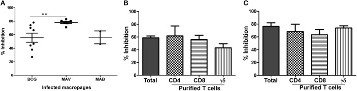
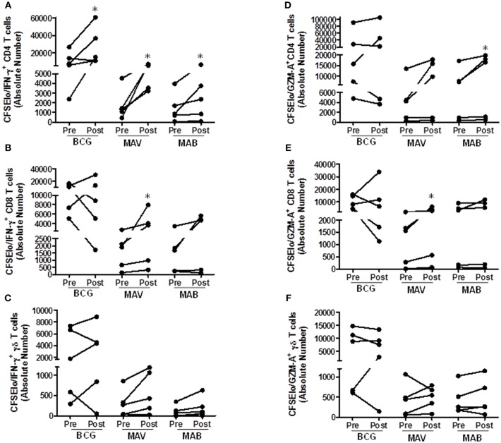
Pulmonary non-tuberculous mycobacterial (NTM) infections particularly caused by Mycobacterium avium complex (MAC) and Mycobacterium abscessus (MAB) are becoming major health problems in the U.S. New therapies or vaccines which will help prevent the disease, shorten treatment duration and/or increase treatment success rates are urgently needed. This study was conducted with the objective of testing the hypothesis that Bacillus Calmette Guerin (BCG), a vaccine used for prevention of serious forms of tuberculosis (TB) in children and adolescents in tuberculosis hyperendemic countries, induces cross-protective T cell immunity against Mycobacterium avium (MAV) and MAB. Human TB and NTM cross-protective T cells were quantified using flow cytometric assays. The ability of BCG expanded T cells to inhibit the intracellular growth of MAV and MAB was assessed in co-cultures with infected autologous macrophages. In both BCG-vaccinated and M. tuberculosis (Mtb)-infected mice, NTM cross-reactive immunity was measured using IFN-γ ELISPOT assays. Our results demonstrate the following key findings: (i) peripheral blood mononuclear cells from TB skin test-positive individuals contain MAV and MAB cross-reactive T cells, (ii) both BCG vaccination and Mtb infection of mice induce MAV and MAB cross-reactive splenic cells, (iii) BCG-expanded T cells inhibit intracellular MAV and MAB, (iv) CD4, CD8, and γδ T cells play important roles in inhibition of intracellular MAV and MAB and (v) BCG vaccination of healthy volunteers induces TB and NTM cross-reactive T cells. In conclusion, BCG-vaccination induces NTM cross-reactive immunity, and has the potential for use as a vaccine or immunotherapy to prevent and/or treat pulmonary NTM disease.

Keywords: BCG; abscessus; avium; mycobacteria; nontuberculous.


Figures

Figure 1
Figure 1
Previous exposure to BCG or Mtb induces cross-reactive T cells against NTM. PBMC from BCG-vaccinated and/or latent TB infected individuals (n = 6) were CFSE-labeled and stimulated with optimal concentrations of live BCG (Connaught) or MAV (ATCC 700898). On day 7, cells were restimulated with PMA/ionomycin and the total percentages of CFSElo (proliferating) and IFN-γ producing T cells were determined by flow cytometry. (A) Flow cytometry plots of a single volunteer. Lymphocytes gated on the basis of forward and side scatter and then regated on CD3+ cells were analyzed for CFSE and IFN-γ expression by use of FlowJo software. The number in the upper left quadrant of each dot plot refers to the percentage of CFSEloIFN-γ+CD3+T cells detected after in vitro stimulation. (B) Composite data from 6 volunteers. Stimulation indices were calculated by dividing the absolute numbers of CFSEloIFN-γ+CD3+ T cells in cultures containing BCG or MAV by the absolute numbers of CFSElo/IFN-γ+ CD3+T cells in medium-rested cultures. Stimulation with MAV led to expansion of effector T cells comparable to the level obtained with BCG stimulation (P > 0.05, Wilcoxon matched-pairs test). The bars show ranges and the lines show mean values. NS, not significant.
Figure 2
Figure 2
MAV and MAB cross-reactive immunity includes Th1 and Th17 responses. (A) Schematic of experiments conducted to measure MAV or MAB cross-reactive immunity. PBMC from BCG-vaccinated or latent TB infected individuals (n = 6) were stimulated with BCG or rested in media for 7 days. Then, these expanded cells were co-cultured with MAV- or MAB-infected autologous monocytes at an E:T ratio of 10:1. On day 3 of co-culture, Th1, Th2, and Th17 responses were measured in co-culture supernatants using CBA. Fold changes for each cytokine was calculated by dividing the amount of cytokine produced following restimulation with MAV or MAB by the amount produced in medium-rested cultures. (B) Exposure of BCG-expanded T cells to MAV-infected macrophages increased IL-17, IFN-γ, and TNF-α by 156 ± 62, 11 ± 1.7, 10.3 ± 2.9 pg/ml (Mean ± SE), respectively. IL-10, IL-6, and IL-2 showed no marked changes with fold changes (mean ± SE) of 0.3 ± 0.1, 0.9 ± 0.2, 0.9 ± 0.2 pg/ml, respectively. (C) Similarly, exposure of BCG-expanded T cells to MAB-infected macrophages increased IL-17 and IFN-γ in by 7.2 ± 1.6, and 5.6 ± 2, mean fold ± SE. There were no marked changes in the levels of TNF-α, IL-10, IL-6, and IL-2 with fold changes (mean ± SE) of 1.2 ± 0.3, 0.2 ± 0.2, 0.6 ± 0.3, 0.8 ± 0.4 pg/ml, respectively.
Figure 3
Figure 3
BCG-specific T cells cross-protect against MAV and MAB. Total PBMC or subsets of T cells purified after 7 days of optimal BCG stimulation were co-cultured with autologous macrophages infected with MAV or MAB (E:T of 10). Residual mycobacteria quantified 3 days after co-culture and percent inhibition calculated by dividing the number of residual mycobacteria in the presence BCG-stimulated PBMC by the number of residual mycobacteria in control cultures containing medium-rested PBMC. (A) BCG-expanded T cells inhibit intracellular MAV (n = 8) and MAB (n = 5) as potently as they inhibit intracellular BCG (n = 8). BCG-expanded T cells inhibited intracellular MAV better than intracellular BCG (**p < 0.01, Mann-Whitney U test). (B) Pure CD4, CD8, and γδ T cells inhibited intracellular MAV, and the level of inhibition was similar to inhibition by total BCG-expanded PBMC. (C) Pure CD4, CD8, and γδ T cells inhibited intracellular MAB, and the level of inhibition is similar to the levels of inhibition mediated by total BCG-expanded PBMC.
Figure 4
Figure 4
BCG vaccination or Mtb infection of mice induces MAV and MAB reactive immunity. (A) Groups of C57BL/6 mice (n = 4–5 per group) were vaccinated once or twice intranasally with BCG. Four weeks later mice were euthanized. Splenic cells were harvested from vaccinated and control mice. Cells were rested in medium or stimulated overnight with live BCG at MOI of 3, MAV at MOI of 3, and MAV-WL in IFN-γ ELISPOT assays. Shown are the means ± SE of IFN-γ SFC per million splenic cells. The number of IFN-γ SFC following stimulation with BCG, MAV, and MAV-WL were significantly higher in mice which received one or two BCG vaccinations compared to unvaccinated mice (*P < 0.05, Mann-Whitney U test). The number of mycobacteria-induced IFN-γ SFC were similar following one versus two BCG vaccination (P > 0.05). (B) C57BL6 infected with aerosolized Mtb (n = 5) were euthanized 4 weeks after infection. Splenic cells from uninfected and infected mice were used in IFN-γ ELISPOT assays. Mtb-infected mice had significantly more MAV and MAB cross-reactive IFN-γ SFC compared to uninfected mice (*P < 0.05, Mann-Whitney U test).
Figure 5
Figure 5
BCG vaccination in humans induces MAV and MAB cross-reactive T cells. Paired pre-and post-vaccination PBMC from recently BCG vaccinated volunteers living in St. Louis, MO (n = 5) were used. PBMC were labeled with CFSE and stimulated with BCG, MAB WL, or MAB WL. Medium rested PBMC were used as negative controls. On day 7, cells were restimulated with PMA/ionomycin for 2 h, viable cells were counted and cells were stained for surface and intracellular markers for flow cytometry study. (A–C) show the data for proliferating and IFN-γ producing T cells. (D–F) show the data for proliferating and GZM-A producing T cells. There were significantly higher absolute numbers (AN, per ml of cultures) of BCG-reactive CFSEloIFN-γ+CD4+ T cells (P = 0.03, Wilcoxon Matched Pairs test), MAV WL reactive CFSEloIFN-γ+CD4+ T cells (P = 0.03), MAV WL reactive CFSEloIFN-γ+CD8+ T cells (P = 0.03), MAV WL reactive CFSEloGranzyme A+CD8+ T cells (P = 0.03), MAB WL reactive CFSEloIFN-γ+CD4+ T cells (P = 0.03), and MAB WL reactive CFSEloGranzyme A+CD4+ T cells (P = 0.03), indicating that BCG induces NTM cross-reactive immunity. *P < 0.05.

Similar articles

Cited by

References

    1. Iseman MD, Marras TK. The importance of nontuberculous mycobacterial lung disease. Am J Respir Crit Care Med. (2008) 178:999–1000. 10.1164/rccm.200808-1258ED - DOI - PubMed
    1. Adjemian J, Olivier KN, Seitz AE, Holland SM, Prevots DR. Prevalence of nontuberculous mycobacterial lung disease in U.S. Medicare beneficiaries. Am J Respir Crit Care Med. (2012) 185:881–6. 10.1164/rccm.201111-2016OC - DOI - PMC - PubMed
    1. Cassidy PM, Hedberg K, Saulson A, McNelly E, Winthrop KL. Nontuberculous mycobacterial disease prevalence and risk factors: a changing epidemiology. Clin Infect Dis. (2009) 49:e124–9. 10.1086/648443 - DOI - PubMed
    1. Mirsaeidi M, Machado RF, Garcia JG, Schraufnagel DE. Nontuberculous mycobacterial disease mortality in the United States, 1999-2010: a population-based comparative study. PLoS ONE (2014) 9:e91879. 10.1371/journal.pone.0091879 - DOI - PMC - PubMed
    1. Strollo SE, Adjemian J, Adjemian MK, Prevots DR. The burden of pulmonary nontuberculous mycobacterial disease in the United States. Ann Am Thorac Soc. (2015) 12:1458–64. 10.1513/AnnalsATS.201503-173OC - DOI - PMC - PubMed

Publication types

MeSH terms