Review
. 2019 Feb 19:10:128.
doi: 10.3389/fpls.2019.00128. eCollection 2019.

Identifying and Engineering Genes for Parthenogenesis in Plants

Affiliations
Review

Identifying and Engineering Genes for Parthenogenesis in Plants

Kitty Vijverberg et al. Front Plant Sci. .

Abstract

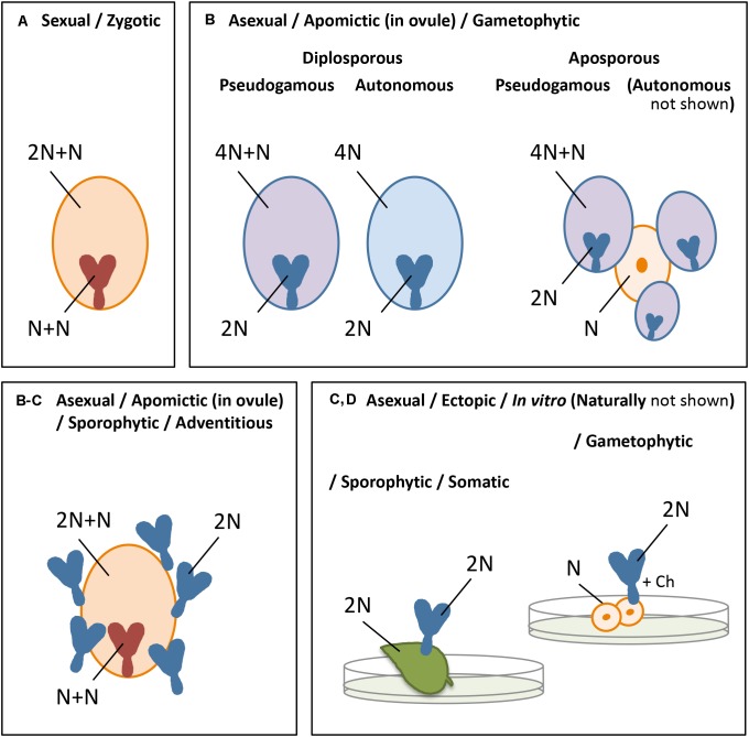
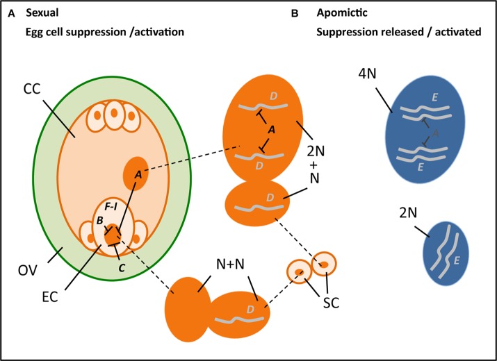
Parthenogenesis is the spontaneous development of an embryo from an unfertilized egg cell. It naturally occurs in a variety of plant and animal species. In plants, parthenogenesis usually is found in combination with apomeiosis (the omission of meiosis) and pseudogamous or autonomous (with or without central cell fertilization) endosperm formation, together known as apomixis (clonal seed production). The initiation of embryogenesis in vivo and in vitro has high potential in plant breeding methods, particularly for the instant production of homozygous lines from haploid gametes [doubled haploids (DHs)], the maintenance of vigorous F1-hybrids through clonal seed production after combining it with apomeiosis, reverse breeding approaches, and for linking diploid and polyploid gene pools. Because of this large interest, efforts to identify gene(s) for parthenogenesis from natural apomicts have been undertaken by using map-based cloning strategies and comparative gene expression studies. In addition, engineering parthenogenesis in sexual model species has been investigated via mutagenesis and gain-of-function strategies. These efforts have started to pay off, particularly by the isolation of the PsASGR-BabyBoom-Like from apomictic Pennisetum, a gene proven to be transferable to and functional in sexual pearl millet, rice, and maize. This review aims to summarize the current knowledge on parthenogenesis, the possible gene candidates also outside the grasses, and the use of these genes in plant breeding protocols. It shows that parthenogenesis is able to inherit and function independently from apomeiosis and endosperm formation, is expressed and active in the egg cell, and can induce embryogenesis in polyploid, diploid as well as haploid egg cells in plants. It also shows the importance of genes involved in the suppression of transcription and modifications thereof at one hand, and in embryogenesis for which transcription is allowed or artificially overexpressed on the other, in parthenogenetic reproduction. Finally, it emphasizes the importance of functional endosperm to allow for successful embryo growth and viable seed production.

Keywords: Pennisetum; PsASGR-BabyBoom-Like (PsASGR-BBML); Taraxacum; apomixis; doubled haploids; embryo induction; embryogenesis; parthenogenesis.


Figures

FIGURE 1
FIGURE 1
Summary of the different forms of embryogenesis in plants, showing the embryo and endosperm originated from a mature embryo sac (A,B) or the embryo ectopically (C,D) after sexual (A) or asexual (B–D) reproduction, with orange indicating the sexual process, blue the asexual or apomictic process, pink apomictic reproduction with fertilization of the central cell, and N = chromosome set after reduction division: (A) zygotic embryogenesis, involving chromosome reduction (N) and gamete fusion (N+N for the embryo, 2N+N for the endosperm), (B) apomictic embryogenesis, occurring in the ovule, either gametophytic apomixis in which an embryo sac arises from an unreduced megaspore (diplospory) or sporophytic cell of the ovule, usually adjacent to a sexually derived spore or developing embryo sac (apospory), and parthenogenetic (spontaneous) embryo development and autonomous (spontaneous) or pseudogamous (after fertilization of the central cell) endosperm formation, or sporophytic apomixis in which the embryo arises directly from a sporophytic cell of the ovule, often as polyembryony and alongside a sexually derived embryo and endosperm (C) somatic/sporophytic embryogenesis, involving ectopic embryo development from sporophytic cells, and (D) gametophytic embryogenesis, idem from a gametophytic cell. The latter two (C,D) omit the formation of an embryo sac, endosperm, and a seed coat, and occur naturally, for example, from leaf margins or ovular cells (C), gametophytic tissue in lower plants or, e.g., a synergid (D), but are particularly known from in vitro embryogenesis in which embryos are formed in culture, after external induction, particularly from protoplasts, leaf, the hypocotyl or other plant tissues (C), or microspores (D).
FIGURE 2
FIGURE 2
Hypothetical cartoon summarizing the potential processes and genes involved in parthenogenesis (A–I, with ---| denoting suppression) (see also Table 1), with left an ovule (OV, green) that encloses a reduced embryo sac (orange), including an egg cell (EC), central cell (CC), two synergids (next to the EC), and three antipodal cells, and outside it two sperm cells (SC, orange), in the middle the enlarged nuclei (dark orange) of the central cell (top, 2N; N = chromosome set after reduction division), sperm cells (middle, each N) and egg cell (bottom, N) at karyogamy, and right the unreduced nuclei (dark blue) of an apomictic central cell (top, 4N) and egg cell (bottom, 2N). A, B, and C denote genes involved in the suppression of transcription in the egg cell, particularly present in the central cell (A) notably the FIS-PRC2, egg cell (B) possibly RKD-TF, and the surrounding ovular tissue (C) potentially AGO9. D and E denote genes that support embryogenesis either after fertilization and transferring a paternally expressed, maternally silenced gene (D) as hypothesized for Os-BBM1, or via natural expression in an apomict (E) as is found for PsASGR-BBML and possibly involves reduced or absence of suppression. FI denote genes and other stimuli that are found to be involved in embryogenesis either after ectopic overexpression to induce somatic embryogenesis (F), for example, LEC2, increase embryogenesis capacity (G), e.g., SERK1, being involved in auxin response (H) as recently found for bHLH49, or changes in physiological conditions and abiotic stress factors (I) such as Ca2+ and heat stress.

References

    1. Agashe B., Prasad C. K., Siddiqi I. (2002). Identification and analysis of DYAD: a gene required for meiotic chromosome organisation and female meiotic progression in Arabidopsis. Development 129 3935–3943. - PubMed
    1. Akiyama Y., Conner J. A., Goel S., Morishige D. T., Mullet J. E., Hanna W. W., et al. (2004). High-resolution physical mapping in Pennisetum squamulatum reveals extensive chromosomal heteromorphism of the genomic region associated with apomixis. Plant Physiol. 134 1733–1741. 10.1104/pp.103.033969 - DOI - PMC - PubMed
    1. Albertini E. A., Porceddu F., Ferranti L., Reale G., Barcaccia B., Romano B., et al. (2001). Apospory and parthenogenesis may be uncoupled in Poa pratensis: a cytological investigation. Sex. Plant Reprod. 14 213–217. 10.1007/s00497-001-0116-2 - DOI - PubMed
    1. Anderson S. N., Johnson C. S., Chesnut J., Jones D. S., Khanday I., Woodhouse M., et al. (2017). The zygotic transition is initiated in unicellular plant zygotes with asymmetric activation of parental genomes. Dev. Cell 43 349–358. 10.1016/j.devcel.2017.10.005 - DOI - PubMed
    1. Anderson S. N., Johnson C. S., Jones D. S., Conrad L. J., Gou X., Russell S. D., et al. (2013). Transcriptomes of isolated Oryza sativa gametes characterized by deep sequencing: evidence for distinct sex-dependent chromatin and epigenetic states before fertilization. Plant J. 76 729–741. 10.1111/tpj.12336 - DOI - PubMed

LinkOut - more resources