. 2019 Jan 29:2019:4265040.
doi: 10.1155/2019/4265040. eCollection 2019.

Downregulation of PTPRK Promotes Cell Proliferation and Metastasis of NSCLC by Enhancing STAT3 Activation

Affiliations

Downregulation of PTPRK Promotes Cell Proliferation and Metastasis of NSCLC by Enhancing STAT3 Activation

Xuting Xu et al. Anal Cell Pathol (Amst). .

Abstract

Objective: The receptor-type tyrosine-protein phosphatase κ (PTPRK) is a candidate tumor suppressor involved in the tumorigenesis of various organs. However, its expression and biological roles in non-small-cell lung cancer (NSCLC) have not yet been investigated.

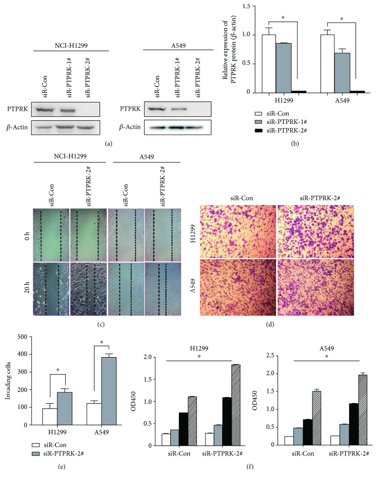
Methods: PTPRK expression in NSCLC tissues and cell lines was examined using real-time PCR and western blotting. In addition, the effects of PTPRK on cell migration, invasion, and proliferation were evaluated in vitro. Furthermore, we explored whether the downregulation of PTPRK led to STAT3 activation in NSCLC cell lines by western blotting. The expression of phospho-STAT3Tyr705 in primary human NSCLC tissues was evaluated by immunohistochemistry.

Results: The results showed that PTPRK expression was frequently reduced in NSCLC tissues with lymph node metastasis and cell lines. The inhibition of PTPRK expression resulted in increased proliferation, invasion, and migration of NSCLC cells in vitro. Additionally, after silencing of PTPRK, phospho-STAT3Tyr705 was significantly increased in NSCLC cells. Moreover, the phospho-STAT3Tyr705 levels of NSCLC tissues were positively correlated with lymph node metastasis and significantly inversely correlated with the expression of PTPRK (p < 0.05).

Conclusions: These results suggested that PTPRK functions as a novel tumor suppressor in NSCLC, and its suppressive ability may be involved in STAT3 activation.


Figures

Figure 1
Figure 1
PTPRK is frequently underexpressed in NSCLC with lymph node (LN) metastasis. (a) PTPRK mRNA expression was quantified by qRT-PCR in 30 lung tumors with non-lymph node metastasis and 16 tumors with lymph node metastasis. (b) qRT-PCR analysis of PTPRK expression levels in one normal human bronchial epithelial cell (16HBE) and seven NSCLC cell lines, and expression levels were all normalized to 16HBE.
Figure 2
Figure 2
PTPRK knockdown promotes the cell proliferation, migration, and invasion ability in H1299 and A549 cells. (a) Western blotting analysis protein H1299 and A549 cells transfected two chemically synthesized siRNAs. (b) Quantitative analysis of PTPRK protein levels was calibrated with beta-actin levels of each sample from (a). (c) Representative micrographs of wound healing assay of the H1299 and A549 cells transfected with PTPRK siRNA#2 or NC. Wound closures were photographed at 0 h and 20 h after wounding. (d) Representative micrographs of Transwell invasion assay of the H1299 and A549 cells transfected with PTPRK siRNA#2 or NC. (e) Quantification of indicated invading cells in five random fields analyzed by the Transwell assays. Values represent the mean ± SD from three independent measurements. (f) Cell proliferation assays. H1299 and A549 cells were transfected with PTPRK siRNA#2 or NC. Cells were counted by a CCK-8 kit after 6 h, 24 h, 48 h, and 72 h. Values represent the mean ± SD from three independent measurements.
Figure 3
Figure 3
PTPRK downregulation contributes to STAT3 activation and is associated with poor prognosis of NSCLC. (a) The protein expression level of phopho-STAT3Tyr705, STAT3, and PTPRK was measured by western blotting in H1299 cells transfected with NC and PTPRK siRNA#2. (b) Densitometric quantifications of phopho-STAT3Tyr705, STAT3, and PTPRK protein levels in H1299 cells transfected with NC and PTPRK siRNA#2 according to (a). (c) Representative micrographs of immunohistochemical staining of the phopho-STAT3Tyr705 protein (brown nuclear staining) in 26 NSCLC tissues. (A) negative control; (B) weak positive (+) expression, weak staining pattern; (C) positive expression (++), medium staining pattern; (D) strong positive expression (+++), strong staining pattern. (d) Correlation between phopho-STAT3Tyr705 and PTPRK expression was analyzed. Expression of PTPRK in 26 clinical tissue samples was measured by real-time PCR.

References

    1. Torre L. A., Bray F., Siegel R. L., Ferlay J., Lortet-Tieulent J., Jemal A. Global cancer statistics, 2012. CA: a Cancer Journal for Clinicians. 2015;65(2):87–108. doi: 10.3322/caac.21262. - DOI - PubMed
    1. Suzuki A., Suzuki M., Mizushima-Sugano J., et al. Sequencing and phasing cancer mutations in lung cancers using a long-read portable sequencer. DNA Research. 2017;24(6):585–596. doi: 10.1093/dnares/dsx027. - DOI - PMC - PubMed
    1. Spring K., Fournier P., Lapointe L., et al. The protein tyrosine phosphatase DEP-1/PTPRJ promotes breast cancer cell invasion and metastasis. Oncogene. 2015;34(44):5536–5547. doi: 10.1038/onc.2015.9. - DOI - PubMed
    1. Stoker A. W. Protein tyrosine phosphatases and signalling. Journal of Endocrinology. 2005;185(1):19–33. doi: 10.1677/joe.1.06069. - DOI - PubMed
    1. Starr T. K., Allaei R., Silverstein K. A. T., et al. A transposon-based genetic screen in mice identifies genes altered in colorectal cancer. Science. 2009;323(5922):1747–1750. doi: 10.1126/science.1163040. - DOI - PMC - PubMed

MeSH terms

Substances

LinkOut - more resources