Review
. 2019 Feb 8;6(2):104-117.
doi: 10.1002/mdc3.12730. eCollection 2019 Feb.

Resting-state Functional MRI in Parkinsonian Syndromes

Affiliations
Review

Resting-state Functional MRI in Parkinsonian Syndromes

Massimo Filippi et al. Mov Disord Clin Pract. .

Abstract

Background: Functional MRI (fMRI) has been widely used to study abnormal patterns of functional connectivity at rest in patients with movement disorders such as idiopathic Parkinson's disease (PD) and atypical parkinsonisms.

Methods: This manuscript provides an educational review of the current use of resting-state fMRI in the field of parkinsonian syndromes.

Results: Resting-state fMRI studies have improved the current knowledge about the mechanisms underlying motor and non-motor symptom development and progression in movement disorders. Even if its inclusion in clinical practice is still far away, resting-state fMRI has the potential to be a promising biomarker for early disease detection and prediction. It may also aid in differential diagnosis and monitoring brain responses to therapeutic agents and neurorehabilitation strategies in different movement disorders.

Conclusions: There is urgent need to identify and validate prodromal biomarkers in PD patients, to perform further studies assessing both overlapping and disease-specific fMRI abnormalities among parkinsonian syndromes, and to continue technical advances to fully realize the potential of fMRI as a tool to monitor the efficacy of chronic therapies.

Keywords: Parkinson's disease (PD); atypical parkinsonisms; functional MRI (fMRI); magnetic resonance imaging (MRI); resting‐state.


Figures

Figure 1
Figure 1
Resting‐state brain networks representation and functions.
Figure 2
Figure 2
Main motor symptoms‐related (a) and cognitive symptoms‐related (b) functional connectivity alterations in PD patients.
Figure 3
Figure 3
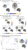
Resting‐state functional MRI in multiple system atrophy (MSA). (A) Significant differences in functional connectivity within the pontocerebellar (left panel) and default mode networks (right) in all MSA patients compared with healthy controls. Voxelwise t‐tests demonstrated significantly higher functional connectivity in the pons and cerebellar tonsils within the pontocerebellar network and lower functional connectivity along the long‐distance midline cores of the default mode network. (B) Subgroup analysis in MSA‐C and MSA‐P subtypes compared with controls: ANOVA indicated a significant difference in the pontocerebellar (left upper panel) and default mode network (right upper panel). Post‐hoc testing indicated pronounced differences in the cerebellar network in MSA‐C versus controls (left lower panel), whereas lower functional coupling in the default mode network (right lower panel) was more prominent in MSA‐P versus controls. (C) Correlation analysis in all MSA patients with characteristic smooth pursuit impairment demonstrated significant correlations between higher functional connectivity and the shape of saccadized smooth pursuit. (A, B, C) Resulting P‐values were corrected at a 5% FDR‐level with further cluster‐wise correction. Reproduced with permission from Rosskopf et al.92
Figure 4
Figure 4
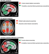
Maps of functional connectivity obtained from the dentate nucleus of healthy controls (red‐yellow) (one‐sample t‐test, P < 0.05, corrected for family wise error [FWE]; upper panel). Maps of differences in the functional connectivity of the dentate nucleus between patients with progressive supranuclear palsy syndrome (PSP; decreased connectivity in blue) and healthy controls, and between patients with corticobasal syndrome (CBS) and healthy controls (increased connectivity in red) (lower panels). Results are obtained using two‐sample t‐test, P < 0.05, corrected for FWE. Abbreviations: L, left hemisphere; R, right hemisphere. Reproduced with permission from Rosskopf et al.98

References

    1. Agosta F, Gatti R, Sarasso E, et al. Brain plasticity in Parkinson's disease with freezing of gait induced by action observation training. J Neurol 2017;264(1):88–101. - PubMed
    1. Lehericy S, Vaillancourt DE, Seppi K, et al. The role of high‐field magnetic resonance imaging in parkinsonian disorders: Pushing the boundaries forward. Mov Disord 2017;32(4):510–525. - PubMed
    1. Heim B, Krismer F, De Marzi R, Seppi K. Magnetic resonance imaging for the diagnosis of Parkinson's disease. J Neural Transm 2017;124(8):915–964. - PMC - PubMed
    1. Coleman P, Federoff H, Kurlan R. A focus on the synapse for neuroprotection in Alzheimer disease and other dementias. Neurology 2004;63(7):1155–1162. - PubMed
    1. Hacker CD, Perlmutter JS, Criswell SR, Ances BM, Snyder AZ. Resting‐state functional connectivity of the striatum in Parkinson's disease. Brain 2012;135(Pt 12):3699–3711. - PMC - PubMed

LinkOut - more resources