. 2019 Jan;19(1):11-21.
doi: 10.1177/1535759718822847. Epub 2019 Jan 31.

The Insula and Its Epilepsies

Affiliations

The Insula and Its Epilepsies

Barbara C Jobst et al. Epilepsy Curr. 2019 Jan.

Abstract

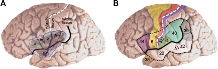
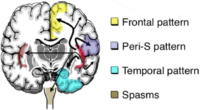
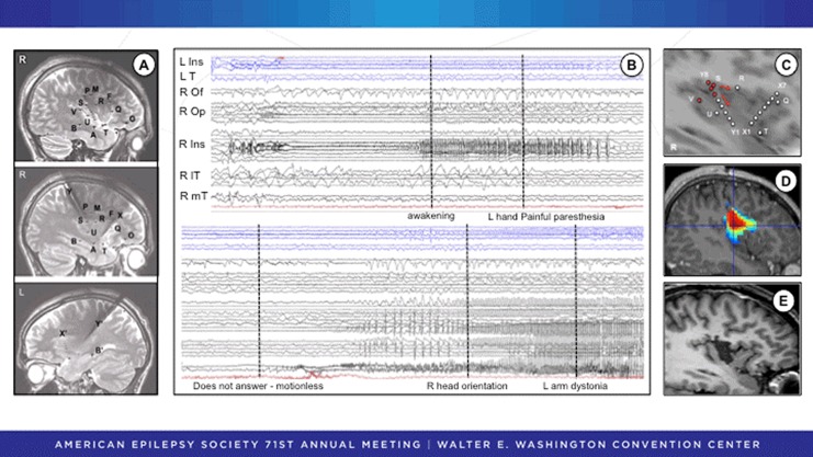
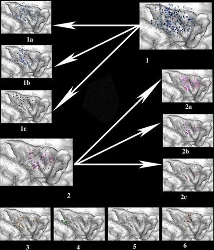
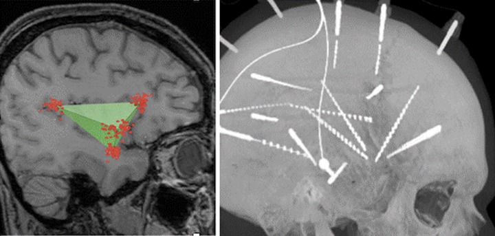
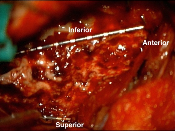
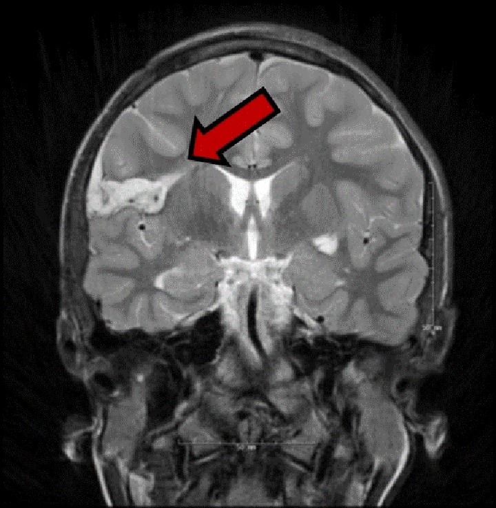
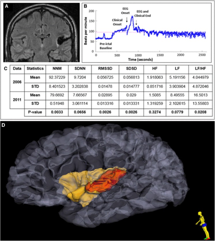
Insular seizures are great mimickers of seizures originating elsewhere in the brain. The insula is a highly connected brain structure. Seizures may only become clinically evident after ictal activity propagates out of the insula with semiology that reflects the propagation pattern. Insular seizures with perisylvian spread, for example, manifest first as throat constriction, followed next by perioral and hemisensory symptoms, and then by unilateral motor symptoms. On the other hand, insular seizures may spread instead to the temporal and frontal lobes and present like seizures originating from these regions. Due to the location of the insula deep in the brain, interictal and ictal scalp electroencephalogram (EEG) changes can be variable and misleading. Magnetic resonance imaging, magnetic resonance spectroscopy, magnetoencephalography, positron emission tomography, and single-photon computed tomography imaging may assist in establishing a diagnosis of insular epilepsy. Intracranial EEG recordings from within the insula, using stereo-EEG or depth electrode techniques, can prove insular seizure origin. Seizure onset, most commonly seen as low-voltage, fast gamma activity, however, can be highly localized and easily missed if the insula is only sparsely sampled. Moreover, seizure spread to the contralateral insula and other brain regions may occur rapidly. Extensive sampling of the insula with multiple electrode trajectories is necessary to avoid these pitfalls. Understanding the functional organization of the insula is helpful when interpreting the semiology produced by insular seizures. Electrical stimulation mapping around the central sulcus of the insula results in paresthesias, while stimulation of the posterior insula typically produces painful sensations. Visceral sensations are the next most common result of insular stimulation. Treatment of insular epilepsy is evolving, but poses challenges. Surgical resections of the insula are effective but risk significant morbidity if not carefully planned. Neurostimulation is an emerging option for treatment, especially for seizures with onset in the posterior insula. The close association of the insula with marked autonomic changes has led to interest in the role of the insula in sudden unexpected death in epilepsy and warrants additional study with larger patient cohorts.


Conflict of interest statement

Declaration of Conflicting Interests: The author(s) declared no potential conflicts of interest with respect to the research, authorship, and/or publication of this article.

Figures

Figure 1.
Figure 1.
A, The insula is anatomically subdivided in an anterior part that comprises 3 short gyri (a, anterior; m, middle; p, posterior) and a posterior part that comprises 2 long gyri (A, anterior; P, posterior). B, The insula is covered by the suprasylvian and infrasylvian opercular regions that are essential for motor, sensory, auditory, and language processing. Numbers refer to Brodmann’s area. CS indicates central sulcus of the insula.
Figure 2.
Figure 2.
Various spread patterns of insula seizures to symptomatogenic zones. Peri-S indicates perisylvian.
Figure 3.
Figure 3.
SEEG recording of a patient with very localized insular onset and subsequent insular resection. A, The SEEG study was focused on the right insulo-opercular region with additional electrodes sampling the right temporal and frontal lobes and the left insular and temporal regions. B, SEEG activity at seizure onset (upper panel) exhibits spikes and polyspike discharges quickly followed by a low-voltage fast activity in the superior part of the anterior long gyrus of the right insula (R Ins) that spreads to the right opercular cortex (R Op). Note the almost immediate involvement of the contralateral insula (L Ins). The patient describes a painful tingling sensation in the left hand and then (lower panel) loses contact and presents temporal-like symptoms when the seizure spread to the right mesiotemporal lobe (R mT), anteroinferior part of the insula (R Ins), and lateral temporal cortex (lT). The orbitofrontal cortex (Of) is spared. C, Schematic representation of the insular contacts involved in seizure onset before spreading to extrainsular regions. D, Epileptogenicity map indicating the highest value of activation in the 60- to 100-Hz frequency band at seizure onset. E, Tailored resection of the right anterior long insular gyrus. Postoperatively, there was transient dysgeusia that resolved completely. The patient has been seizure free without medication for 6 years. Pathological examination revealed a focal cortical dysplasia type IB. SEEG indicates stereo electroencephalogram.
Figure 4.
Figure 4.
Location and type of symptoms evoked by electrical stimulations of the insular cortex. (1) Somatosensory responses, including nonpainful, nonthermal sensations (light blue, 1A), thermal sensations (medium blue, 1B), and painful sensations (deep blue, 1C). (2) Visceral sensations, including constrictive sensations (light pink, 2A), viscero-vegetative sensations (deep pink, 2B), and viscero-psychic symptoms ( pink, 2C). (3) Vestibular sensations (orange). (4) Auditory sensations (green). (5) Speech disturbances (violet). (6) Olfactogustatory sensations (red for taste, yellow for smell).
Figure 5.
Figure 5.
The insula is shaped like a tetrahedron or triangular pyramid (left). By taking oblique approaches to the insula and implanting 4 electrodes, we are able to mimic this anatomy and define the borders of the insula (right).
Figure 6.
Figure 6.
Intraoperative view of SEEG electrodes serving as internal landmarks during insular resection. A complete anterior insulectomy has been performed, as superior, inferior, and anterior insular electrodes can be seen at the borders of the resection. SEEG indicates stereo electroencephalogram.
Figure 7.
Figure 7.
Postoperative T2 MRI image (coronal orientation) after right caudal rostral insula resection in a 22-year-old female, resulting in symptomatic infarct in the ipsilateral corona radiata (red arrow). The procedure resulted in seizure freedom, but with a only partially recovered left side hemiparesis. MRI indicates magnetic resonance imaging.
Figure 8.
Figure 8.
A, Postoperative MRI FLAIR sequence shows evidence of a left posterior temporo-insular resection cavity with surrounding gliosis in a patient with later SUDEP. B, Heart rate plots show ictal sinus tachycardia, followed by sustained postictal sinus tachycardia lasting at least 25 minutes after a nonfatal generalized convulsive seizure. C, Heart rate time and frequency domain parameters calculated during the presurgery (2006) and postsurgery (2011) epilepsy monitoring unit (EMU) evaluations and the results from generalized estimating equation (GEE) analysis. D Extent of insular resection and damage, after 3-dimensional reconstruction of pre- and postoperative MRI is delineated in red. MRI indicates magnetic resonance imaging; FLAIR, fluid-attenuated inversion recovery; SUDEP, sudden unexpected death in epilepsy; MNN, mean of normal to normal heart beats; SDNN, standard deviation of normal to normal heart beats.

References

    1. Morel A, Gallay MN, Baechler A, Wyss M, Gallay DS. The human insula: architectonic organization and postmortem MRI registration. Neuroscience. 2013;236:117–135. - PubMed
    1. Kurth F, Zilles K, Fox PT, Laird AR, Eickhoff SB. A link between the systems: functional differentiation and integration within the human insula revealed by meta-analysis. Brain Struct Funct. 2010;214(4-5):519–534. - PMC - PubMed
    1. Collin G, Sporns O, Mandl RC, van den Heuvel MP. Structural and functional aspects relating to cost and benefit of rich club organization in the human cerebral cortex. Cerebral Cortex. 2014;24(9):2258–2267. - PMC - PubMed
    1. Namkung H, Kim SH, Sawa A. The insula: an underestimated brain area in clinical neuroscience, psychiatry, and neurology. Trends Neurosci. 2017;40(4):200–207. - PMC - PubMed
    1. Dimova P. The insula: semiology In: Arzimanoglou A, Cross JH, Gaillard WD, et al. (Eds.), Pediatric Epilepsy Surgery. Montrouge, France: John Libbey Eurotext; 2016:121–129.

LinkOut - more resources