. 2019 May;130(5):804-819.
doi: 10.1097/ALN.0000000000002648.

α2δ-1-Bound N-Methyl-D-aspartate Receptors Mediate Morphine-induced Hyperalgesia and Analgesic Tolerance by Potentiating Glutamatergic Input in Rodents

Affiliations

α2δ-1-Bound N-Methyl-D-aspartate Receptors Mediate Morphine-induced Hyperalgesia and Analgesic Tolerance by Potentiating Glutamatergic Input in Rodents

Meichun Deng et al. Anesthesiology. 2019 May.

Abstract

Background: Chronic use of μ-opioid receptor agonists paradoxically causes both hyperalgesia and the loss of analgesic efficacy. Opioid treatment increases presynaptic N-methyl-D-aspartate receptor activity to potentiate nociceptive input to spinal dorsal horn neurons. However, the mechanism responsible for this opioid-induced activation of presynaptic N-methyl-D-aspartate receptors remains unclear. α2δ-1, formerly known as a calcium channel subunit, interacts with N-methyl-D-aspartate receptors and is primarily expressed at presynaptic terminals. This study tested the hypothesis that α2δ-1-bound N-methyl-D-aspartate receptors contribute to presynaptic N-methyl-D-aspartate receptor hyperactivity associated with opioid-induced hyperalgesia and analgesic tolerance.

Methods: Rats (5 mg/kg) and wild-type and α2δ-1-knockout mice (10 mg/kg) were treated intraperitoneally with morphine twice/day for 8 consecutive days, and nociceptive thresholds were examined. Presynaptic N-methyl-D-aspartate receptor activity was recorded in spinal cord slices. Coimmunoprecipitation was performed to examine protein-protein interactions.

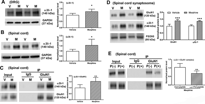
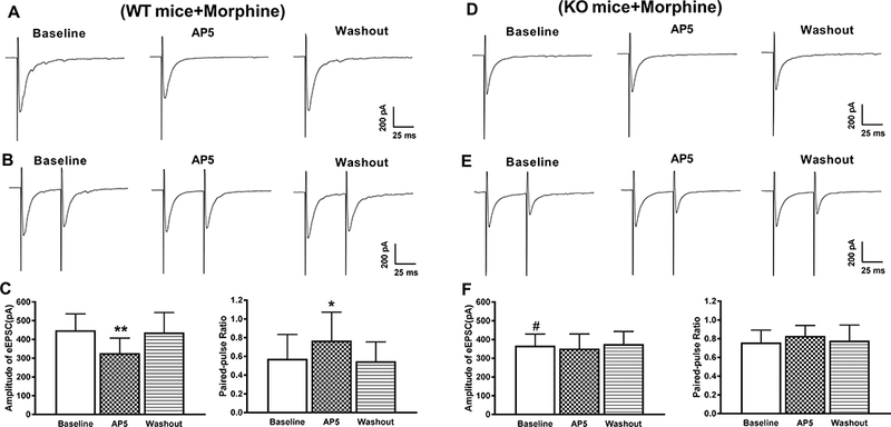
Results: Chronic morphine treatment in rats increased α2δ-1 protein amounts in the dorsal root ganglion and spinal cord. Chronic morphine exposure also increased the physical interaction between α2δ-1 and N-methyl-D-aspartate receptors by 1.5 ± 0.3 fold (means ± SD, P = 0.009, n = 6) and the prevalence of α2δ-1-bound N-methyl-D-aspartate receptors at spinal cord synapses. Inhibiting α2δ-1 with gabapentin or genetic knockout of α2δ-1 abolished the increase in presynaptic N-methyl-D-aspartate receptor activity in the spinal dorsal horn induced by morphine treatment. Furthermore, uncoupling the α2δ-1-N-methyl-D-aspartate receptor interaction with an α2δ-1 C terminus-interfering peptide fully reversed morphine-induced tonic activation of N-methyl-D-aspartate receptors at the central terminal of primary afferents. Finally, intraperitoneal injection of gabapentin or intrathecal injection of an α2δ-1 C terminus-interfering peptide or α2δ-1 genetic knockout abolished the mechanical and thermal hyperalgesia induced by chronic morphine exposure and largely preserved morphine's analgesic effect during 8 days of morphine treatment.

Conclusions: α2δ-1-Bound N-methyl-D-aspartate receptors contribute to opioid-induced hyperalgesia and tolerance by augmenting presynaptic N-methyl-D-aspartate receptor expression and activity at the spinal cord level.


Conflict of interest statement

Conflicts of Interest

The authors declare no competing interests with the contents of this study.

Figures

Figure 1.
Figure 1.. Chronic morphine treatment increases α2δ-1 association with NMDARs at spinal cord synapses.
A and B, Representative blots and quantification of α2δ-1 protein levels in the DRG (A) and dorsal spinal cord (B) from vehicle-treated (V) and morphine-treated (M) rats (n = 6 rats in each group). C, Coimmunoprecipitation analysis shows that GluN1 coprecipitated with α2δ-1 in the membrane extracts of dorsal spinal cord tissues of rats treated with vehicle or morphine for 8 days (n = 6 rats in each group). The amount of α2δ-1 proteins was normalized to that of GluN1 in the same sample, and the mean α2δ-1 level in vehicle-treated rats was considered to be 1. D, Representative gel images and quantification of GluN1 and α2δ-1 protein amounts in dorsal spinal cord synaptosomes from vehicle- and morphine-treated rats (n = 6 rats in each group). E, Coimmunoprecipitation analysis shows the effect of treatment with 1 μM α2δ-1Tat peptide and scrambled control peptide on the α2δ-1-GluN1 complex level in spinal cord slices from morphine-treated rats (n = 6 rats in each group). Data are shown as means ± SD. *P < 0.05, **P < 0.01, ***P < 0.001 vs. the vehicle or control peptide group.
Figure 2.
Figure 2.. α2δ-1 mediates chronic morphine exposure-induced potentiation of presynaptic NMDAR activity in the spinal dorsal horn.
A, Representative recording traces and cumulative plots show the effect of bath application of 50 μM AP5 on the frequency and amplitude of mEPSCs of a lamina II neuron from a morphine-treated rat. B, Summary data show the effect of 50 μM AP5 on the mean frequency and amplitude of mEPSCs of lamina II neurons (n = 11 neurons) from morphine-treated rats. C, Representative recording traces and cumulative plots show that bath application of 50 μM AP5 had no effect on the frequency or amplitude of mEPSCs of a lamina II neuron pretreated with 100 μM gabapentin from a morphine-treated rat. D, Summary data show no effect from 50 μM AP5 on the mean frequency or amplitude of mEPSCs of lamina II neurons (n = 10 neurons) pretreated with 100 μM gabapentin from morphine-treated rats. Data are shown as means ± SD. **P < 0.01 vs. the baseline. ###P < 0.001 vs. the baseline in the morphine + vehicle group.
Figure 3.
Figure 3.. α2δ-1 is involved in chronic morphine exposure-induced potentiation of NMDAR activity at primary afferent terminals in the spinal dorsal horn.
A and B, Representative recording traces show the effect of bath application of 50 μM AP5 on evoked monosynaptic EPSCs (A) and the paired-pulse ratio (PPR, B) of a vehicle-incubated lamina II neuron from a morphine-treated rat. C, Summary data show the effect of 50 μM AP5 on the amplitude (n = 10 neurons) and PPR (n = 10 neurons) of evoked monosynaptic EPSCs of vehicle-incubated lamina II neurons in morphine-treated rats. D and E, Representative recording traces show no effect from bath application of 50 μM AP5 on the amplitude of monosynaptically evoked EPSCs (D) or the PPR (E) of a lamina II neuron in spinal cord slices pretreated with 100 μM gabapentin in a morphine-treated rat. F, Summary data show no effect from 50 μM AP5 on the mean amplitude (n = 11 neurons) or PPR (n = 11 neurons) of monosynaptic EPSCs of lamina II neurons pretreated with 100 μM gabapentin from morphine-treated rats. Data are shown as means ± SD. *P < 0.05, ***P < 0.001 vs. the baseline. ##P < 0.01 vs. the baseline in the morphine + vehicle group.
Figure 4.
Figure 4.. α2δ-1 is essential for the chronic morphine exposure-induced activation of presynaptic NMDARs in the spinal dorsal horn.
A, Representative recording trace and cumulative plots show the effect of bath application of 50 μM AP5 on the frequency and amplitude of mEPSCs of a lamina II neuron from a morphine-treated WT mouse. B, Summary data show the effect of 50 μM AP5 on the mean frequency and amplitude of mEPSCs (n = 11 neurons) in spinal cord slices from morphine-treated WT mice. C, Representative recording traces and cumulative plots show no effect from AP5 on the frequency or amplitude of mEPSCs of a lamina II neuron from a morphine-treated α2δ-1 KO mouse. D, Summary data show no effect of AP5 on the mean frequency or amplitude of mEPSCs (n = 10 neurons) in spinal cord slices from morphine-treated α2δ-1 KO mice. Data are shown as means ± SD. ***P < 0.001 vs. the baseline. ##P < 0.01 vs. the baseline in the WT group.
Figure 5.
Figure 5.. α2δ-1 is required for the chronic morphine exposure-induced increase in NMDAR activity at primary afferent terminals.
A and B, Representative current traces show the effect of bath application of 50 μM AP5 on the amplitude of monosynaptic EPSCs (A) and the PPR (B) of a lamina II neuron from a morphine-treated WT mouse. C, Summary data show the effect of 50 μM AP5 on the mean amplitude (n = 11 neurons) and PPR (n = 11 neurons) of monosynaptic EPSCs of lamina II neurons from spinal cord slices of morphine-treated WT mice. C and D, Representative current traces show no effect of AP5 on the mean amplitude of evoked monosynaptic EPSCs (C) or PPR (D) of a lamina II neuron of a morphine-treated α2δ-1 KO mouse. E, Group data show the lack of effect of 50 μM AP5 on the amplitude (n = 11 neurons) and the PPR (n = 11 neurons) of monosynaptic EPSCs of lamina II neurons from spinal cord slices of morphine-treated α2δ-1 KO mice. Data are shown as means ± SD. **P < 0.05, **P < 0.01 vs. the baseline. #P < 0.05 vs. the baseline in the WT group.
Figure 6.
Figure 6.. α2δ-1–bound NMDARs mediate the chronic morphine exposure-induced increase in presynaptic NMDAR activity in the spinal cord.
A, Representative recording traces and cumulative plots show the effect of bath application of 50 μM AP5 on the frequency and amplitude of mEPSCs of a lamina II neuron pretreated with control peptide (1 μM) from a spinal cord slice of a morphine-treated rat. B, Summary data show the effect of 50 μM AP5 on the mean frequency and amplitude of mEPSCs (n = 10 neurons) in spinal cord slices pretreated with control peptide from morphine-treated rats. C, Representative recording traces and cumulative plots show no effect of AP5 on the frequency or amplitude of mEPSCs of a lamina II neuron pretreated with α2δ-1Tat peptide (1 μM) from a spinal cord slice of a morphine-treated rat. D, Summary data show no effect of AP5 on the mean frequency or amplitude of mEPSCs (n = 11 neurons) in spinal cord slices pretreated with α2δ-1Tat peptide from morphine-treated rats. Data are shown as means ± SD. ***P < 0.001 vs. the baseline. ###P < 0.001 vs. the baseline in the morphine + control peptide group.
Figure 7.
Figure 7.. α2δ-1–bound NMDARs are critically involved in chronic morphine exposure-induced activation of NMDARs at primary afferent terminals.
A and B, Representative current traces show the effect of bath application of 50 μM AP5 on the amplitude of monosynaptic EPSCs (A) and the PPR (B) of a lamina II neuron from a spinal cord slice pretreated with control peptide (1 μM) from a morphine-treated rat. C, Summary data show the effect of 50 μM AP5 on the mean amplitude (n = 11 neurons) and PPR (n = 11 neurons) of monosynaptic EPSCs of lamina II neurons from spinal cord slices pretreated with control peptide from morphine-treated rats. C and D, Representative current traces show no effect of AP5 on the amplitude of evoked monosynaptic EPSCs (C) or PPR (D) of a lamina II neuron from a spinal cord slice pretreated with α2δ-1Tat peptide (1 μM) from a morphine-treated rat. E, Summary data show no effect of AP5 on the mean amplitude (n = 11 neurons) or PPR (n = 11 neurons) of monosynaptic EPSCs of lamina II neurons from spinal cord slices pretreated with α2δ-1Tat peptide from morphine-treated rats. Data are shown as means ± SD. **P < 0.01, ***P < 0.001 vs. the baseline. ###P < 0.001 vs. the baseline in the morphine + control peptide group.
Figure 8.
Figure 8.. α2δ-1 at the spinal cord level mediates chronic morphine exposure-induced hyperalgesia and analgesic tolerance.
A and B, Time course of changes in the baseline mechanical (A) and thermal (B) withdrawal thresholds and the analgesic effect of morphine in rats treated with systemic morphine plus vehicle (n = 8 rats) or gabapentin (100 mg/kg, n = 8 rats). C and D, Time course of changes in the baseline mechanical (C) and thermal (D) withdrawal thresholds and the analgesic effect of morphine in rats treated with systemic morphine plus control peptide (1 μg) or α2δ-1Tat peptide (1 μg) (n = 10 rats in each group). E and F, Time course of changes in the baseline mechanical (E) and thermal (F) withdrawal thresholds and the analgesic effect of morphine in WT and α2δ-1 KO mice (n = 8 mice per group). The baseline withdrawal threshold was measured before each morphine injection, and the analgesic effect of morphine was tested 30 min after morphine injection. *P < 0.05, **P < 0.01, ***P < 0.001 vs. values at day 1. Data are shown as means ± SD. #P < 0.05, ##P < 0.01, ###P < 0.001 vs. values in the corresponding control group (vehicle, control peptide, or WT) at the same time point.
Figure 9.
Figure 9.. Treatment with gabapentin or α2δ-1Tat peptide attenuates established hyperalgesia and analgesic tolerance induced by chronic morphine treatment.
A and B, Effect of intraperitoneal (i.p.) injection of vehicle or gabapentin (100 mg/kg, i.p.) on the baseline mechanical (A) and heat (B) withdrawal thresholds and the acute analgesic effect of morphine (5 mg/kg, i.p.) in rats pretreated with chronic morphine for 8 days (n = 8 rats in each groups). C and D, Effect of intrathecal injection of α2δ-1Tat peptide (1 μg) or control peptide (1 μg) on the baseline mechanical (C) and heat (D) withdrawal thresholds and the acute analgesic effect of morphine (5 mg/kg, i.p.) in rats pretreated with chronic morphine for 8 days (n = 8 rats in each group). BL, pre-morphine treatment baseline. Data are shown as means ± SD. *P < 0.05, **P < 0.01, ***P < 0.001 vs. values at time 0. #P < 0.05, ###P < 0.001 vs. corresponding values in the vehicle or control peptide group at the same time point.

Similar articles

Cited by

References

    1. Ferrini F, Trang T, Mattioli TAM, Laffray S, Del’Guidice T, Lorenzo LE, Castonguay A, Doyon N, Zhang WB, Godin AG, Mohr D, Beggs S, Vandal K, Beaulieu JM, Cahill CM, Salter MW, De Koninck Y: Morphine hyperalgesia gated through microglia-mediated disruption of neuronal Cl- homeostasis. Nat Neurosci 2013; 16: 183–192 - PMC - PubMed
    1. Chen SR, Pan HL: Loss of TRPV1-expressing sensory neurons reduces spinal mu opioid receptors but paradoxically potentiates opioid analgesia. J Neurophysiol 2006; 95: 3086–96 - PubMed
    1. Chen SR, Pan HL: Blocking mu opioid receptors in the spinal cord prevents the analgesic action by subsequent systemic opioids. Brain Res 2006; 1081: 119–25 - PubMed
    1. Zhao YL, Chen SR, Chen H, Pan HL: Chronic opioid potentiates presynaptic but impairs postsynaptic N-methyl-D-aspartic acid receptor activity in spinal cords: implications for opioid hyperalgesia and tolerance. J Biol Chem 2012; 287: 25073–85 - PMC - PubMed
    1. Corder G, Tawfik VL, Wang D, Sypek EI, Low SA, Dickinson JR, Sotoudeh C, Clark JD, Barres BA, Bohlen CJ, Scherrer G: Loss of mu opioid receptor signaling in nociceptors, but not microglia, abrogates morphine tolerance without disrupting analgesia. Nat Med 2017; 23: 164–173 - PMC - PubMed

Publication types