. 2018 Mar 23:7:17162.
doi: 10.1038/lsa.2017.162. eCollection 2018.

X-ray focusing with efficient high-NA multilayer Laue lenses

Affiliations

X-ray focusing with efficient high-NA multilayer Laue lenses

Saša Bajt et al. Light Sci Appl. .

Abstract

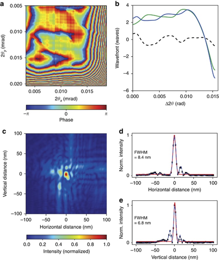
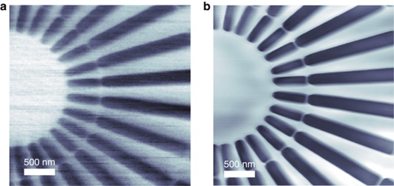
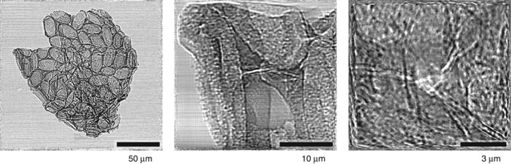
Multilayer Laue lenses are volume diffraction elements for the efficient focusing of X-rays. With a new manufacturing technique that we introduced, it is possible to fabricate lenses of sufficiently high numerical aperture (NA) to achieve focal spot sizes below 10 nm. The alternating layers of the materials that form the lens must span a broad range of thicknesses on the nanometer scale to achieve the necessary range of X-ray deflection angles required to achieve a high NA. This poses a challenge to both the accuracy of the deposition process and the control of the materials properties, which often vary with layer thickness. We introduced a new pair of materials-tungsten carbide and silicon carbide-to prepare layered structures with smooth and sharp interfaces and with no material phase transitions that hampered the manufacture of previous lenses. Using a pair of multilayer Laue lenses (MLLs) fabricated from this system, we achieved a two-dimensional focus of 8.4 × 6.8 nm2 at a photon energy of 16.3 keV with high diffraction efficiency and demonstrated scanning-based imaging of samples with a resolution well below 10 nm. The high NA also allowed projection holographic imaging with strong phase contrast over a large range of magnifications. An error analysis indicates the possibility of achieving 1 nm focusing.

Keywords: X-ray holography; X-ray optics; multilayer Laue lenses; multilayers; ptychography.


Conflict of interest statement

The authors declare no conflict of interest.

Figures

Figure 1
Figure 1
To achieve high diffraction efficiency across the entire pupil of a multilayer Laue lens, the layer periods dn at heights hn must follow the zone plate law such that reflected rays constructively interfere at the focus. The layers must be wedged so that Bragg’s law sin θn=λ/(2dn) is satisfied locally at every bi-layer for a wavelength λ. For a lens of focal length f, this places the layers normal to a circle of radius 2f. The lens can be thought of as an off-axis portion of a larger parent lens. The numerical aperture is given by sin θNA, where θNA is half of the difference between the largest and smallest deflection angles.
Figure 2
Figure 2
Plot of the far-field diffracted intensity as a function of the angular position from the W/SiC MLL (a) (adapted from Ref. 16). A localized phase error at a scattering angle of approximately 10.3 mrad at 0.056 nm wavelength from the W/SiC lens gave rise to an obvious intensity spike. At that position, the multilayer period was approximately 5.5 nm. Bright field (left column) and dark field (remaining columns) TEM images of W/SiC (b) and WC/SiC (c) have periods from 4.0 to 10 nm (as described in the Materials and Methods section). The white bar in all images corresponds to 20 nm. The transition from amorphous to crystalline W layers occurs at a period of approximately 5.7 nm.
Figure 3
Figure 3
Experimental setup used at the P11 and HXN beamlines. Two MLLs are orthogonal to each other, as indicated by the red double-headed arrows. At P11, a LAMBDA detector with 55-μm pixels was used to measure the far-field intensity at a distance 1.4 m from the focus. At HXN, a Timepix detector with 55-μm pixels was placed 0.533 m downstream of the focus. An example of the efficiency measurement is shown from P11, with intensities shown on a logarithmic color scale.
Figure 4
Figure 4
Diffraction efficiency of the (a) vertical and (b) horizontal lenses, as determined by the far-field 1D diffraction pattern of each lens at 16.3 keV photon energy, mapped as a function of tilt of the MLL lens. (c) Maps of the relative diffraction efficiency of the two lenses combined, from the far-field 2D diffraction pattern as a function of photon energy, ranging from 15.5 to 17.5 keV. All intensity maps are shown on a linear color scale. The side of the square pupil corresponds to 20 mrad (at 16.3 keV), and it scales inversely with photon energy.
Figure 5
Figure 5
(a) Wavefront error in the pupil plane of the MLL showing significant error at the edges of the lenses. (b) The unwrapped wavefront separated into 1D phase profiles of each individual lens, and the differences in the phase error of the two lenses (dashed line) indicate an upper limit of the manufacturing reproducibility. (c) Reconstructed intensity in the MLL focus as determined by ptychography. Lineouts in horizontal (d) and vertical directions (e) of the in-focus intensities (black dots) were fitted with Gaussian functions (red lines) with widths of 8.4 and 6.8 nm, respectively. The color bars in (a) and (c) indicate the normalized phase and intensity.
Figure 6
Figure 6
The incoherent STXM absorption-contrast image (a) and ptychography reconstructed image (b) showing a Siemens star with 100 nm inner spikes and a 20 nm spacing ring between the inner and outer spikes.
Figure 7
Figure 7
In-line projection holograms of an Acantharian cyst obtained at the PETRA III P11 beamline using 16.3 keV X-rays. The magnification is increased as the sample is moved towards the X-ray focus. The exposure time of the first image was 2 s, over which time 1.7 × 10⁹ photons were recorded. The exposure time of the other images was 5 s, with 3.7 × 10⁹ detected photons.

References

    1. Ice GE, Budai JD, Pang JWL. The race to x-ray microbeam and nanobeam science. Science 2011; 334: 1234–1239. - PubMed
    1. Maser J, Schmahl G. Coupled wave description of the diffraction by zone plates with high aspect ratios. Opt Common 1992; 89: 355–362.
    1. Yan HF, Conley R, Bouet N, Chu YS. Hard x-ray nanofocusing by multilayer Laue lenses. J Phys D Appl Phys 2014; 47: 263001.
    1. Yan YF, Maser J, Macrander A, Shen Q, Vogt S et al. Takagi-Taupin description of x-ray dynamical diffraction from diffractive optics with large numerical aperture. Phys Rev B 2007; 76: 115438.
    1. Bajt S, Chapman HN, Aquila A, Gullikson E. High-efficiency x-ray gratings with asymmetric-cut multilayers. J Opt Soc Am A 2012; 29: 216–230. - PubMed